

")


转诊至印度金奈市专科医疗中心的COVID-19患者肺血管和胃肠道血管病变的放射学评估:前瞻性的横断面研究
- 作者: Sathishkumar H.1, Faizal A.2, Majith A.2, Raj V.2, Venkataramani A.3
-
隶属关系:
- Government Chengalpattu Medical College and Hospital
- Saveetha Medical College and Hospital, Saveetha Institute of Medical and Technical Sciences
- Mahatma Gandhi Medical College and Research Institute
- 期: 卷 5, 编号 3 (2024)
- 页面: 480-490
- 栏目: 原创性科研成果
- URL: https://ogarev-online.ru/DD/article/view/310033
- DOI: https://doi.org/10.17816/DD626759
- ID: 310033
如何引用文章
全文:

详细
论证。自2019年出现以来,冠状病毒感染大流行(COVID-19)在全球范围内的发病率和死亡率一直居高不下。 COVID-19主要是一种呼吸系统疾病,但也会影响其他器官系统,包括血管和胃肠道。COVID-19的并发症包括动脉和静脉血栓形成,大量研究表明肺栓塞风险增加。对COVID-19患者的病理解剖显示,有肺血管血栓形成和肠道缺血的病例。
目的 — 评估转诊至印度金奈市专科医疗中心的COVID-19 患者肺血管(尤其是肺栓塞)和胃肠道血管病变的放射学体症。
材料和方法。100名符合研究标准的COVID-19患者接受了肺血管计算机断层扫描和腹部造影剂计算机断层扫描。肺血管和肠道血管变化的评估由一名有5年经验的放射科医生进行。为确定 COVID-19与肺部和肠道血管变化之间关系的显著性,进行了统计分析。
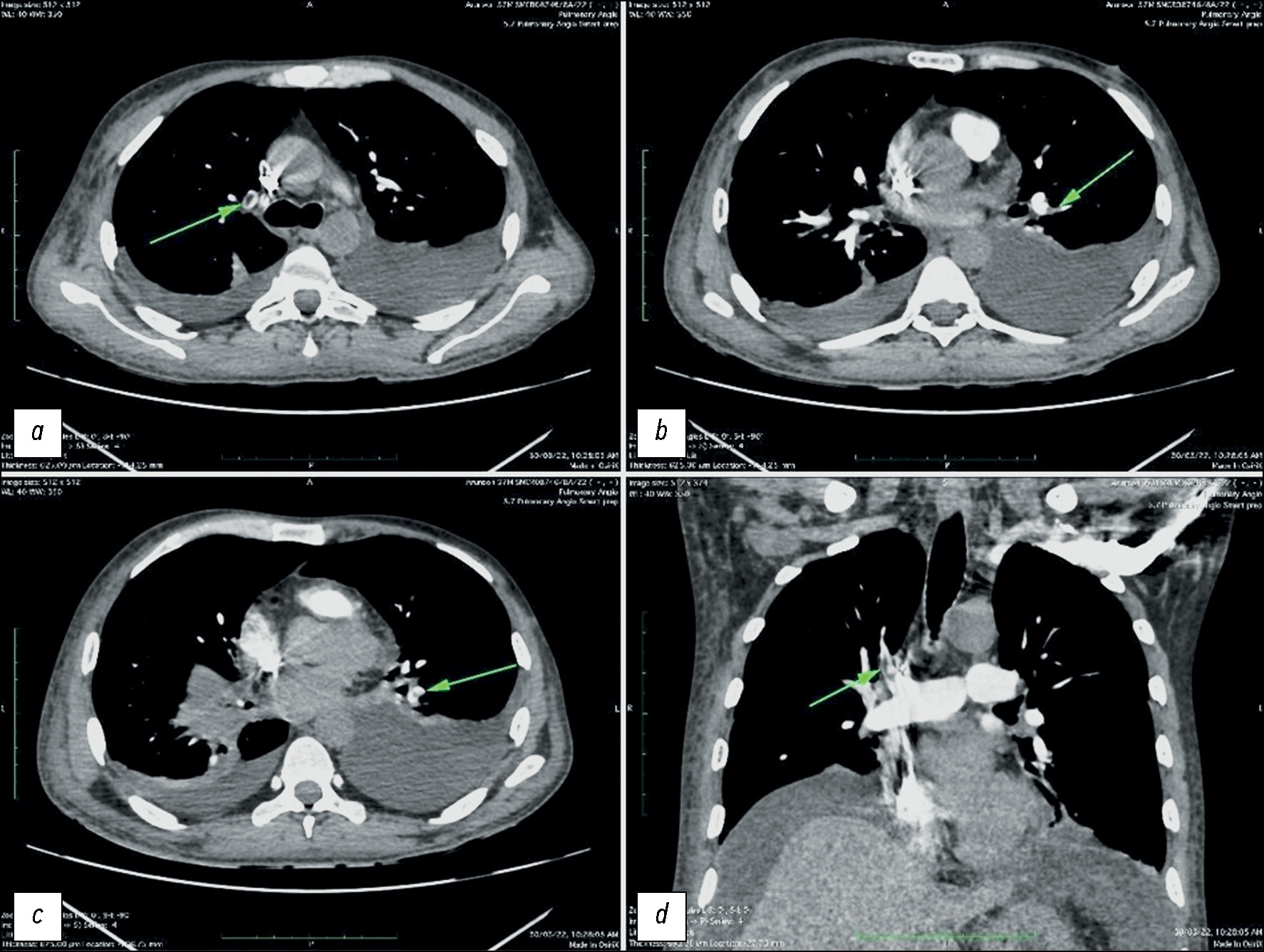
结果。研究中,11 名患者出现肺栓塞,7 名患者的肠道血管出现明显变化,包括肠壁增厚、肠系膜缺血和网膜梗死,这表明 COVID-19患者胃肠道血管潜在的损伤。研究发现,COVID-19的存在与肺栓塞的发生呈正相关性,肺栓塞平均在发病11天后确诊。根据肺血管计算机断层扫描,在24名COVID-19严重急性呼吸道病程的患者中,有7人发现了肺栓塞。此外,在10名接受人工通气的患者中,有7人出现了这种并发症。在7名肠道血管发生变化的患者中,有4人也发现了肺栓塞,这表明这些变化之间存在着显著的关系。观察到的肠道血管变化是由于血管内血栓形成所致。
结论。根据结果可以得知,严重的COVID-19患者往往同时伴有肺栓塞,而且肠道血管也会发生变化。此外,多变量分析显示,有创人工通气与肺栓塞的发生存在关联。因此,这些患者的治疗决策应基于增强计算机断层扫描,因为不使用造影剂的标准计算机断层扫描无法提供必要的信息。
关键词
作者简介
Hariharan Sathishkumar
Government Chengalpattu Medical College and Hospital
Email: harisathish00788@gmail.com
ORCID iD: 0009-0005-3198-9899
MD
印度, ChengalpattuAfwaan Faizal
Saveetha Medical College and Hospital, Saveetha Institute of Medical and Technical Sciences
Email: affanfaizal4498@gmail.com
ORCID iD: 0009-0000-9664-6698
MD
印度, ChennaiAbdul Majith
Saveetha Medical College and Hospital, Saveetha Institute of Medical and Technical Sciences
编辑信件的主要联系方式.
Email: radsaveetha@gmail.com
ORCID iD: 0009-0005-2351-2644
MD
印度, ChennaiVishnu Raj
Saveetha Medical College and Hospital, Saveetha Institute of Medical and Technical Sciences
Email: vishnurajsedhu@gmail.com
ORCID iD: 0009-0004-2436-4586
MD
印度, ChennaiAgathiyanathan Venkataramani
Mahatma Gandhi Medical College and Research Institute
Email: aakashv3@gmail.com
ORCID iD: 0009-0004-7403-8164
印度, Puducherry
参考
- Manfrini N, Notarbartolo S, Grifantini R, Pesce E. SARS-CoV-2: A Glance at the Innate Immune Response Elicited by Infection and Vaccination. Antibodies. 2024;13(1):13. doi: 10.3390/antib13010013
- Thornton GM, Fleck BA, Kroeker E, et al. The impact of heating, ventilation, and air conditioning design features on the transmission of viruses, including the 2019 novel coronavirus: A systematic review of ventilation and coronavirus. PLOS Glob Public Health. 2022;2(7):e0000552. doi: 10.1371/journal.pgph.0000552
- Chen AT, Wang CY, Zhu WL, Chen W. Coagulation Disorders and Thrombosis in COVID-19 Patients and a Possible Mechanism Involving Endothelial Cells: A Review. Aging Dis. 2022;13(1):144–156. doi: 10.14336/AD.2021.0704
- Nemec HM, Ferenczy A, Christie BD 3rd, Ashley DW, Montgomery A. Correlation of D-dimer and Outcomes in COVID-19 Patients. Am Surg. 2022;88(9):2115–2118. doi: 10.1177/00031348221091940
- Stark K, Massberg S. Interplay between inflammation and thrombosis in cardiovascular pathology. Nat Rev Cardiol. 2021;18:666–682. doi: 10.1038/s41569-021-00552-1
- Al-Samkari H, Karp Leaf RS, Dzik WH, et al. COVID-19 and coagulation: bleeding and thrombotic manifestations of SARS-CoV-2 infection. Blood. 2020;136(4):489–500. doi: 10.1182/blood.2020006520
- Covino M, De Matteis G, Polla DAD, et al. Predictors of in-hospital mortality AND death RISK STRATIFICATION among COVID-19 PATIENTS aged ≥80 YEARs OLD. Archives of Gerontology and Geriatrics. 2021;95:104383. doi: 10.1016/j.archger.2021.104383
- Parekh M, Donuru A, Balasubramanya R, Kapur S. Review of the Chest CT Differential Diagnosis of Ground-Glass Opacities in the COVID Era. Radiology. 2020;297(3):E289–E302. doi: 10.1148/radiol.2020202504
- Danzi GB, Loffi M, Galeazzi G, Gherbesi E. Acute pulmonary embolism and COVID-19 pneumonia: a random association? Eur Heart J. 2020;41(19):1858. doi: 10.1093/eurheartj/ehaa254
- Chi G, Lee JJ, Jamil A, et al. Venous thromboembolism among hospitalized patients with COVID-19 undergoing thromboprophylaxis: a systematic review and meta-analysis. J Clin Med. 2020;9(8):2489. doi: 10.3390/jcm9082489
- Grillet F, Behr J, Calame P, Aubry S, Delabrousse E. Acute pulmonary embolism associated with COVID-19 pneumonia detected with pulmonary CT angiography. Radiology. 2020;296(3):E186–E188. doi: 10.1148/radiol.2020201544
- Abdelmohsen MA, Alkandari BM, Gupta VK, et al. Gastrointestinal tract imaging findings in confirmed COVID-19 patients: a non-comparative observational study. Egypt J Radiol Nucl Med. 2021;52. doi: 10.1186/s43055-021-00433-0
补充文件


