

")


基于直肠癌患者初诊分期时获得的肿瘤加权T2核磁共振图像的纹理分析预测新辅助放化疗的效果
- 作者: Dayneko Y.A.1, Berezovskaya T.P.1, Mirzeabasov O.A.2, Starkov S.O.2, Myalina S.A.1, Nevolskikh A.A.1, Ivanov S.А.1,3, Kaprin A.D.3,4,5
-
隶属关系:
- A.F. Tsyb Medical Radiology Research Centre, National Medical Research Radiological Center
- National Research Nuclear University MEPhI (Moscow Engineering Physics Institute)
- Peoples’ Friendship University of Russia
- P.A. Herzen Moscow Research Institute of Oncology, National Medical Research Radiological Center
- National Medical Research Radiological Centre
- 期: 卷 5, 编号 3 (2024)
- 页面: 421-435
- 栏目: 原创性科研成果
- URL: https://ogarev-online.ru/DD/article/view/310028
- DOI: https://doi.org/10.17816/DD628304
- ID: 310028
如何引用文章
全文:

详细
论证。为了预测局部晚期直肠癌对新辅助放化疗的反应,最近人们在寻找潜在无创生物标志物方面一直在做着巨大的努力。
研究目的 — 评估局部晚期直肠癌在原发加权T2图像上的纹理特征,将其作为预测标准新辅助放化疗效果的潜在因素,并在此基础上开发一套预测此类治疗效果的系统。
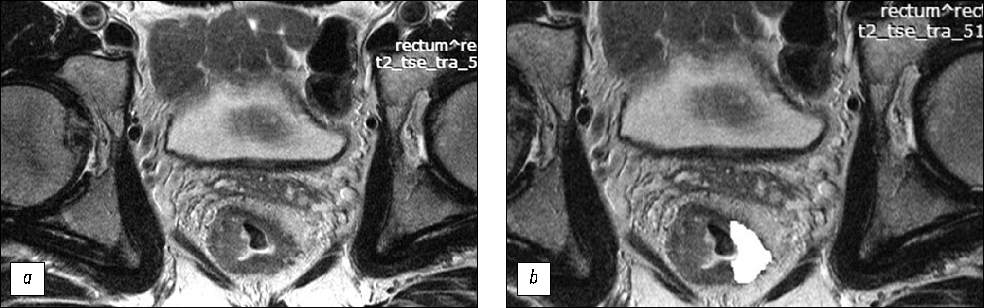
材料和方法。在回顾性研究中接受新辅助放化疗综合治疗的局部晚期直肠癌患者(n=82)被分为训练样本(n=58)和对照样本(n=24)。在肿瘤中心水平使用方向垂直于肠壁的高分辨率原始加权T2图像,用于纹理分析。纹理分析基于灰度级共生矩阵(GLCM),借助MAZDAver计算机程序执行了二阶统计法。 4.6和11个纹理参数的计算。在训练样本中进行手术制剂形态学评估后,查明治疗有反应(预后良好组)和无反应(预后不良组)患者组的纹理分析参数的真实差异,并在此基础上创建评估新辅助放化疗效果的评分系统。系统在对照样本上进行测试确定诊断效率的参数。
结果。在训练样本的预后良好组和预后不良组中找到分离点,其五个纹理参数上存在真实差异:AngScMom(p=0.021)、SumofSqs(p=0.003)、SumEntrp(p=0.003)、Entropy(p=0.038)和 SumVarnc(p=0.015),在创建评分系统时排除了 Entropy,因其与 SumEntrp相比有很强的直接相关性,最低的AUC, 以及与SumofSqs相比重现性低。反应预测评分系统的诊断效率在训练样本中的灵敏度、特异性、阳性预测能力和阴性预测能力分别为 72%、69%、70% 和 71%,相应的在对照样本中分别为 80%、64%、62% 和 82%。曲线下面积在训练样本中为 ROC 为 0.77,在对照样本中为 0.72。
结论。对局部晚期直肠癌患者原发肿瘤T2-VI的纹理分析可以预测诊断效率适中的新辅助放化疗效果,表明这个方向的进一步研究的前景性。
作者简介
Yana A. Dayneko
A.F. Tsyb Medical Radiology Research Centre, National Medical Research Radiological Center
编辑信件的主要联系方式.
Email: vorobeyana@gmail.com
ORCID iD: 0000-0002-4524-0839
SPIN 代码: 1841-7759
MD, Cand. Sci. (Medicine)
俄罗斯联邦, ObninskTatiana P. Berezovskaya
A.F. Tsyb Medical Radiology Research Centre, National Medical Research Radiological Center
Email: tberezovska@yahoo.com
ORCID iD: 0000-0002-3549-4499
SPIN 代码: 5837-3465
MD, Dr. Sci. (Medicine), Professor
俄罗斯联邦, ObninskOleg A. Mirzeabasov
National Research Nuclear University MEPhI (Moscow Engineering Physics Institute)
Email: oami@yandex.ru
ORCID iD: 0000-0001-5587-2795
SPIN 代码: 3820-4320
MD, Assistant Professor
俄罗斯联邦, ObninskSergey O. Starkov
National Research Nuclear University MEPhI (Moscow Engineering Physics Institute)
Email: sergeystarkov56@mail.ru
ORCID iD: 0000-0002-0420-7856
Dr. Sci. (Physical and Mathematical), Professor
俄罗斯联邦, ObninskSofiya A. Myalina
A.F. Tsyb Medical Radiology Research Centre, National Medical Research Radiological Center
Email: samyalina@mail.ru
ORCID iD: 0000-0001-6686-5419
SPIN 代码: 9668-3834
俄罗斯联邦, Obninsk
Aleksey A. Nevolskikh
A.F. Tsyb Medical Radiology Research Centre, National Medical Research Radiological Center
Email: editor@omnidoctor.ru
ORCID iD: 0000-0001-5961-2958
SPIN 代码: 3787-6139
MD, Dr. Sci. (Medicine)
俄罗斯联邦, ObninskSergey А. Ivanov
A.F. Tsyb Medical Radiology Research Centre, National Medical Research Radiological Center; Peoples’ Friendship University of Russia
Email: oncourolog@gmail.com
ORCID iD: 0000-0001-7689-6032
SPIN 代码: 4264-5167
MD, Dr. Sci. (Medicine), Professor, corresponding member of the Russian Academy of Sciences
俄罗斯联邦, Obninsk; MoscowAndrey D. Kaprin
Peoples’ Friendship University of Russia; P.A. Herzen Moscow Research Institute of Oncology, National Medical Research Radiological Center; National Medical Research Radiological Centre
Email: contact@nmicr.ru
ORCID iD: 0000-0001-8784-8415
SPIN 代码: 1759-8101
MD, Dr. Sci. (Medicine), Professor, academician of the Russian Academy of Sciences
俄罗斯联邦, Moscow; Moscow; Moscow参考
- Berdov BA, Erigin DV, Nevolskykh AA, et al. Multidiciplinary approach to the treatment of rectal cancer. Oncology Bulletin Volga region. 2015;(4):21–28. EDN: UKTSNJ
- Maistrenko NA, Galkin VN, Erygin DV, Sazonov AA. Neoadjuvant chemoradiotherapy in combined treatment of patients with rectal cancer. Grekov’s Bulletin Surg. 2017;176(4):31–38. EDN: ZDQHMV doi: 10.24884/0042-4625-2017-176-4-31-38
- Berdov BA, Erygin DV, Nevolskikh AA, et al. Neoadjuvant therapy for locally advanced rectal cancer. P.A. Herzen J Oncology. 2018;3(7):9–15. EDN: XSLIJN doi: 10.17116/onkolog2018739
- Maas M, Nelemans PJ, Valentini V, et al. Long-term outcome in patients with a pathological complete response after chemoradiation for rectal cancer: A pooled analysis of individual patient data. Lancet Oncol. 2010;11(9):835–844. doi: 10.1016/S1470-2045(10)70172-8
- Petresc B, Lebovici A, Caraiani C, et al. Pre-treatment T2-WI based radiomics features for prediction of locally advanced rectal cancer non-response to neoadjuvant chemoradiotherapy: A preliminary study. Cancers (Basel). 2020;12(7):1894. doi: 10.3390/cancers12071894
- Huh JW, Kim HC, Kim SH, et al. Tumor regression grade as a clinically useful outcome predictor in patients with rectal cancer after preoperative chemoradiotherapy. Surgery. 2019;165(3):579–585. doi: 10.1016/j.surg.2018.08.026
- Lambin P, Leijenaar RT, Deist TM, et al. Radiomics: The bridge between medical imaging and personalized medicine. Nat Rev Clin Oncol. 2017;14(12):749–762. doi: 10.1038/nrclinonc.2017.141
- Mayerhoefer ME, Materka A, Langs G, et al. Introduction to radiomics. J Nucl Med. 2020;61(4):488–495. doi: 10.2967/jnumed.118.222893
- Papanikolaou N, Matos C, Koh DM. How to develop a meaningful radiomic signature for clinical use in oncologic patients. Cancer Imaging. 2020;20(1):33. EDN: ROFXND doi: 10.1186/s40644-020-00311-4
- Schick U, Lucia F, Dissaux G, et al. MRI-derived radiomics: Methodology and clinical applications in the field of pelvic oncology. Br J Radiol. 2019;92(1104):20190105. doi: 10.1259/bjr.20190105
- Berezovskaya TP, Dayneko YaA, Nevolskikh AA, et al. A system for evaluating the effectiveness of neoadjuvant chemoradiotherapy in patients with colorectal cancer based on a texture analysis of post-therapeutic t2-wi magnetic resonance imaging. REJR. 2020;10(3):92–101. EDN: DCXHXG doi: 10.21569/2222-7415-2020-10-3-92-101
- Lubner MG, Smith AD, Sandrasegaran K, et al. CT texture analysis: Definitions, applications, biologic correlates, and challenges. Radiographics. 2017;37(5):1483–1503. doi: 10.1148/rg.2017170056
- Rogers W, Thulasi Seetha S, Refaee TA, et al. Radiomics: From qualitative to quantitative imaging. Br J Radiol. 2020;93(1108):20190948. doi: 10.1259/bjr.20190948
- Capobianco E, Dominietto M. From medical imaging to radiomics: Role of data science for advancing precision health. J Pers Med. 2020;10(1):15. doi: 10.3390/jpm10010015
- Dagogo-Jack I, Shaw AT. Tumour heterogeneity and resistance to cancer therapies. Nat Rev Clin Oncol. 2018;15(2):81–94. doi: 10.1038/nrclinonc.2017.166
- Lušnikov EF. Therapeutic pathomorphosis of tumors. In: Kraevskiy NA, Smolyannikova AV, Sarkisova DS, editors. Pathoanatomical diagnosis of human tumors. Moscow: Meditsina; 1993. (In Russ.)
- Miranda J, Horvat N, Assuncao AN, et al. MRI-based radiomic score increased mrTRG accuracy in predicting rectal cancer response to neoadjuvant therapy. AbdomRadiol (NY). 2023;48(6):1911–1920.EDN: IYPGFF doi: 10.1007/s00261-023-03898-x
- Wen L, Liu J, Hu P, et al. MRI-based radiomic models outperform radiologists in predicting pathological complete response to neoadjuvant chemoradiotherapy in locally advanced rectal cancer. AcadRadiol. 2023;30(Suppl. 1):S176–S184. EDN: TYUIWX doi: 10.1016/j.acra.2022.12.037
- Tomaszewski MR, Dominguez-Viqueira W, Ortiz A, et al. Heterogeneity analysis of MRI T2 maps for measurement of early tumor response to radiotherapy. NMR Biomed. 2021;34(3):e4454.EDN: NWLELG doi: 10.1002/nbm.4454
- Stanzione A, Verde F, Romeo V, et al. Radiomics and machine learning applications in rectal cancer: Current update and future perspectives. World J Gastroenterol. 2021;27(32):5306–5321. doi: 10.3748/wjg.v27.i32.5306
- Cui Y, Yang X, Shi Z, et al. Radiomics analysis of multiparametric MRI for prediction of pathological complete response to neoadjuvant chemoradiotherapy in locally advanced rectal cancer. Eur Radiol. 2019;29(3):1211–1220. EDN: ATAEHX doi: 10.1007/s00330-018-5683-9
- Huang H, Han L, Guo J, et al. Multiphase and multiparameter MRI-based radiomics for prediction of tumor response to neoadjuvant therapy in locally advanced rectal cancer. Radiat Oncol. 2023;18(1):179. EDN: ICLFRG doi: 10.1186/s13014-023-02368-4
- Zhou X, Yu Y, Feng Y, et al. Attention mechanism based multi-sequence MRI fusion improves prediction of response to neoadjuvant chemoradiotherapy in locally advanced rectal cancer. Radiat Oncol. 2023;18(1):175. EDN: DIHCZQ doi: 10.1186/s13014-023-02352-y
- Santini D, Danti G, Bicci E, et al. Radiomic features are predictive of response in rectal cancer undergoing therapy. Diagnostics. 2023;13(15):2573. EDN: CWBCMS doi: 10.3390/diagnostics13152573
- Giannini V, Mazzetti S, Bertotto I, et al. Predicting locally advanced rectal cancer response to neoadjuvant therapy with 18F-FDG PET and MRI radiomics features. Eur J Nucl Med Mol Imaging. 2019;46(4):878–888. EDN: PVETLQ doi: 10.1007/s00259-018-4250-6
- Gelezhe PB, Blokhin IA, Semenov SS, Caruso D. Magnetic resonance imaging radiomics in prostate cancer radiology: What is currently known? Digital Diagnostics. 2021;2(4):441−452.EDN: FFFGWI doi: 10.17816/DD70170
- Tibermacine H, Rouanet P, Sbarra M, et al. GRECCAR Study Group. Radiomics modelling in rectal cancer to predict disease-free survival: Evaluation of different approaches. Br J Surg. 2021;108(10):1243–1250. doi: 10.1093/bjs/znab191
- Miranda J, Wang L, Wu X, et al. MRI-based pre-radiomics and delta-radiomics models accurately predict the post-treatment response of rectal adenocarcinoma to neoadjuvant chemoradiotherapy. Front Oncol. 2023;(13):1133008. EDN: XYMVTJ doi: 10.3389/fonc.2023.1133008
- Haralick RM. Statistical and structural approaches to texture. IEEE. 1979;67(5):768–804. doi: 10.1109/PROC.1979.11328
- Mayerhoefer ME, Szomolanyi P, Jirak D, et al. Effects of MRI acquisition parameter variations and protocol heterogeneity on the results of texture analysis and pattern discrimination: An application-oriented study. Med Phys. 2009;36(4):1236–1243. doi: 10.1118/1.3081408
- Shayesteh S, Nazari M, Salahshour A, et al. Treatment response prediction using MRI-based pre-, post-, and delta-radiomic features and machine learning algorithms in colorectal cancer. Med Phys. 2021;48(7):3691–3701. doi: 10.1002/mp.14896
- Song M, Li S, Wang H, et al. MRI radiomics independent of clinical baseline characteristics and neoadjuvant treatment modalities predicts response to neoadjuvant therapy in rectal cancer. Br J Cancer. 2022;127(2):249–257. EDN: BCDXXD doi: 10.1038/s41416-022-01786-7
- Yardimci AH, Kocak B, Sel I, et al. Radiomics of locally advanced rectal cancer: Machine learning-based prediction of response to neoadjuvant chemoradiotherapy using pre-treatment sagittal T2-weighted MRI. JPN J Radiol. 2023;41(1):71–82. EDN: PSFYUW doi: 10.1007/s11604-022-01325-7
补充文件


