

")


一名患有模仿神经母细胞瘤的肾上腺成熟性畸胎瘤的儿童的放射诊断难题
- 作者: Shchelkanova E.S.1, Tereshchenko G.V.1, Krasnov A.S.1
-
隶属关系:
- Dmitry Rogachev National Medical Research Center of Pediatric Hematology, Oncology and Immunology
- 期: 卷 5, 编号 2 (2024)
- 页面: 379-389
- 栏目: 临床病例及临床病例的系列
- URL: https://ogarev-online.ru/DD/article/view/264847
- DOI: https://doi.org/10.17816/DD622768
- ID: 264847
如何引用文章

详细
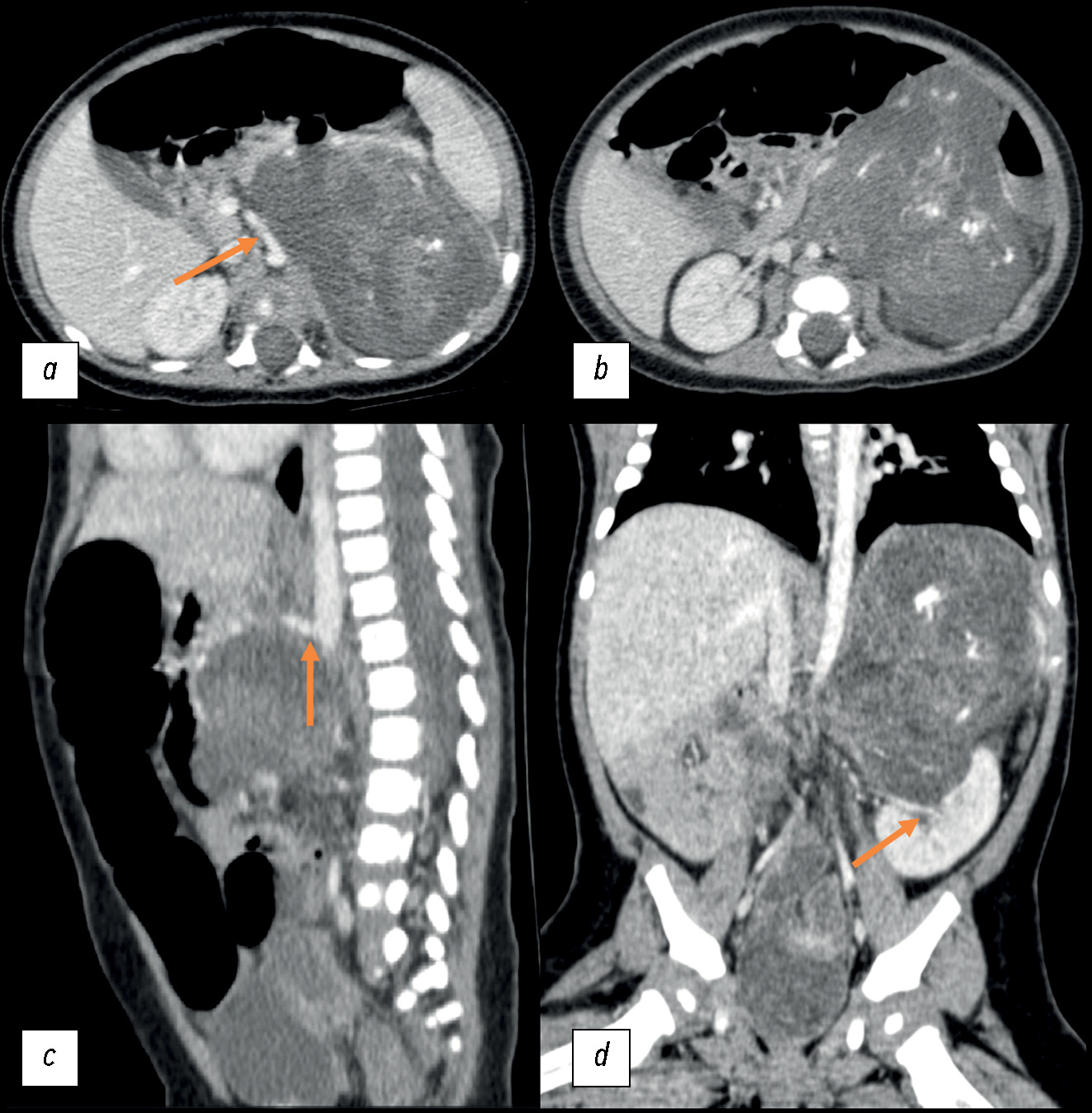
神经母细胞瘤是幼儿最常见的肾上腺肿块,其鉴别系列包括肾母细胞瘤、肾上腺出血、血管肌脂肪瘤、骨髓脂肪瘤和腺瘤。本文描述了一例最罕见的儿童肾上腺肿瘤,即畸胎瘤,尽管其体积较大,但在诊断初期却显示出神经母细胞瘤的所有放射学和组织学特征。
畸胎瘤是生殖细胞肿瘤,通常出现在性腺区域。肾上腺畸胎瘤极为罕见,约占所有肾上腺肿块的 0.13%。肾上腺畸胎瘤通常没有症状,这是因为腹膜后间隙足够大,肿块可以自由生长。
我们首次在俄罗斯文献中介绍了一个 3 个月大儿童肾上腺畸胎瘤的临床病例。文章还详细描述了诊断检查的过程,以及放射科医生和临床医生在罕见部位遇到常见儿童肿瘤时所遇到的困难。
文章旨在帮助医生提高对这种罕见疾病的认识,并将肾上腺畸胎瘤纳入肾上腺肿瘤的潜在鉴别系列。
作者简介
Ekaterina S. Shchelkanova
Dmitry Rogachev National Medical Research Center of Pediatric Hematology, Oncology and Immunology
编辑信件的主要联系方式.
Email: dr.shelkanova@yandex.ru
ORCID iD: 0009-0002-3582-8783
SPIN 代码: 9198-4674
俄罗斯联邦, Moscow
Galina V. Tereshchenko
Dmitry Rogachev National Medical Research Center of Pediatric Hematology, Oncology and Immunology
Email: Galina.Tereshenko@fccho-moscow.ru
ORCID iD: 0000-0001-7317-7104
SPIN 代码: 9413-2500
MD, Cand. Sci. (Medicine)
俄罗斯联邦, MoscowAlexey S. Krasnov
Dmitry Rogachev National Medical Research Center of Pediatric Hematology, Oncology and Immunology
Email: Alexey.Krasnov@fccho-moscow.ru
ORCID iD: 0000-0003-1099-9332
SPIN 代码: 3238-4124
俄罗斯联邦, Moscow
参考
- WHO Classification of Tumours Editorial Board. WHO classification of tumours of endocrine organs, 4th ed. Lloyd R.V., Osamura RY, Kloppel G, Rosai J, editors. Lyon: International Agency for Research on Cancer; 2017.
- Emre Ş, Özcan R, Bakır AC, Kuruğoğlu S, et al. Adrenal masses in children: Imaging, surgical treatment and outcome. Asian J Surg. 2020;43(4):207–212. doi: 10.1016/j.asjsur.2019.03.012
- He C, Yang Y, Yang Y, et al. Teratoma of the adrenal gland: clinical experience and literature review. Gland Surg. 2020;9(4):1056–1064. doi: 10.21037/gs-20-648
- Feoktistova EV, Uskova NG, Varfolomeeva SP, et al. Differential diagnosis of congenital cystic neuroblastoma and prenatal adrenal hemorrhage in children of the first months of life. Pediatric Hematology/Oncology and Immunopathology. 2017;16(1):62–68. doi: 10.24287/1726-1708-2017-16-1-62-68
- Wang X, Li X, Cai H, et al. Rare Primary Adrenal Tumor: A Case Report of Teratomas and Literatures Review. Front Oncol. 2022;12:830003. doi: 10.3389/fonc.2022.830003
- AlQattan A, Alsharit M, Alsaihaty E, et al. The ‘’Monstrous tumor’’ of Adrenal gland: A case report and review of literature on adrenal teratomas. Int. J. Surg. Open. 2023;60:100696. doi: 10.1016/j.ijso.2023.100696
- Craig WD, Fanburg-Smith JC, Henry LR, et al. Fat-containing lesions of the retroperitoneum: radiologic-pathologic correlation. Radiographics. 2009;29(1):261–290. doi: 10.1148/rg.291085203
- Wetherell D, Weerakoon M, Williams D, et al. Mature and Immature Teratoma: A Review of Pathological Characteristics and Treatment Options. Med Surg Urol. 2014;3(1):124. doi: 10.4172/2168-9857.1000124
- Li S, Li H, Ji Z, Yan W, Zhang Y. Primary adrenal teratoma: Clinical characteristics and retroperitoneal laparoscopic resection in five adults. Oncol Lett. 2015;10(5):2865–2870. doi: 10.3892/ol.2015.3701
- Sandoval JA, Williams RF. Neonatal Germ Cell Tumors. Curr Pediatr Rev. 2015;11(3):205–215. doi: 10.2174/1573396311666150714105531
- Wootton-Gorges SL, Thomas KB, Harned RK, et al. Giant cystic abdominal masses in children. Pediatr Radiol. 2005;35(12):1277–1288. doi: 10.1007/s00247-005-1559-7
- Zhao Z, Deng X, Peng L, Kong X. Case Report Management of retroperitoneal teratoma in infants younger than one-year-old. Int J Clin Exp Med. 2018;11(2):1362–1366.
- Singh AP, Jangid M, Morya DP, Gupta A. Retroperitoneal Teratoma in an Infant. Journal of Case Reports. 2014;4(2):317–319. doi: 10.17659/01.2014.0079
- Rattan KN, Kadian YS, Nair VJ, et al. Primary retroperitoneal teratomas in children: a single institution experience. Afr J Paediatr Surg. 2010;7(1):5–8. doi: 10.4103/0189-6725.59350
- Lam AK. Lipomatous tumours in adrenal gland: WHO updates and clinical implications. Endocr Relat Cancer. 2017;24(3):65–79. doi: 10.1530/ERC-16-0564
- Tejedor DC, Gutierrez VR, Afonso JM, et al. Adrenal lipoma: A case report and literature review. Urol Case Rep. 2020;34:101506. doi: 10.1016/j.eucr.2020.101506
- Liao T, Du W, Li X, et al. Recurrent metastatic retroperitoneal dedifferentiated liposarcoma: a case report and literature review. BMC Urol. 2023;23(1):63. doi: 10.1186/s12894-023-01252-3
补充文件


