

")


漏斗胸畸形的磁共振成像
- 作者: Muzafarova G.S.1, Vishnyakova M.V.1, Abramenko A.S.1, Kuzmichev V.A.1, Gatsutsyn V.V.1
-
隶属关系:
- Moscow Regional Research and Clinical Institute
- 期: 卷 5, 编号 2 (2024)
- 页面: 167-177
- 栏目: 原创性科研成果
- URL: https://ogarev-online.ru/DD/article/view/264830
- DOI: https://doi.org/10.17816/DD568087
- ID: 264830
如何引用文章

详细
论证。磁共振成像更常用于确认是否存在漏斗胸畸形,并评估该水平的心脏压迫变化。
目的是通过磁共振成像对漏斗胸畸形进行术前评估。
材料和方法。我们对 38 名患者(30 名男性,8 名女性)的胸部器官磁共振成像进行了回顾性评估。平均年龄为 19.9 岁(±9 岁)。
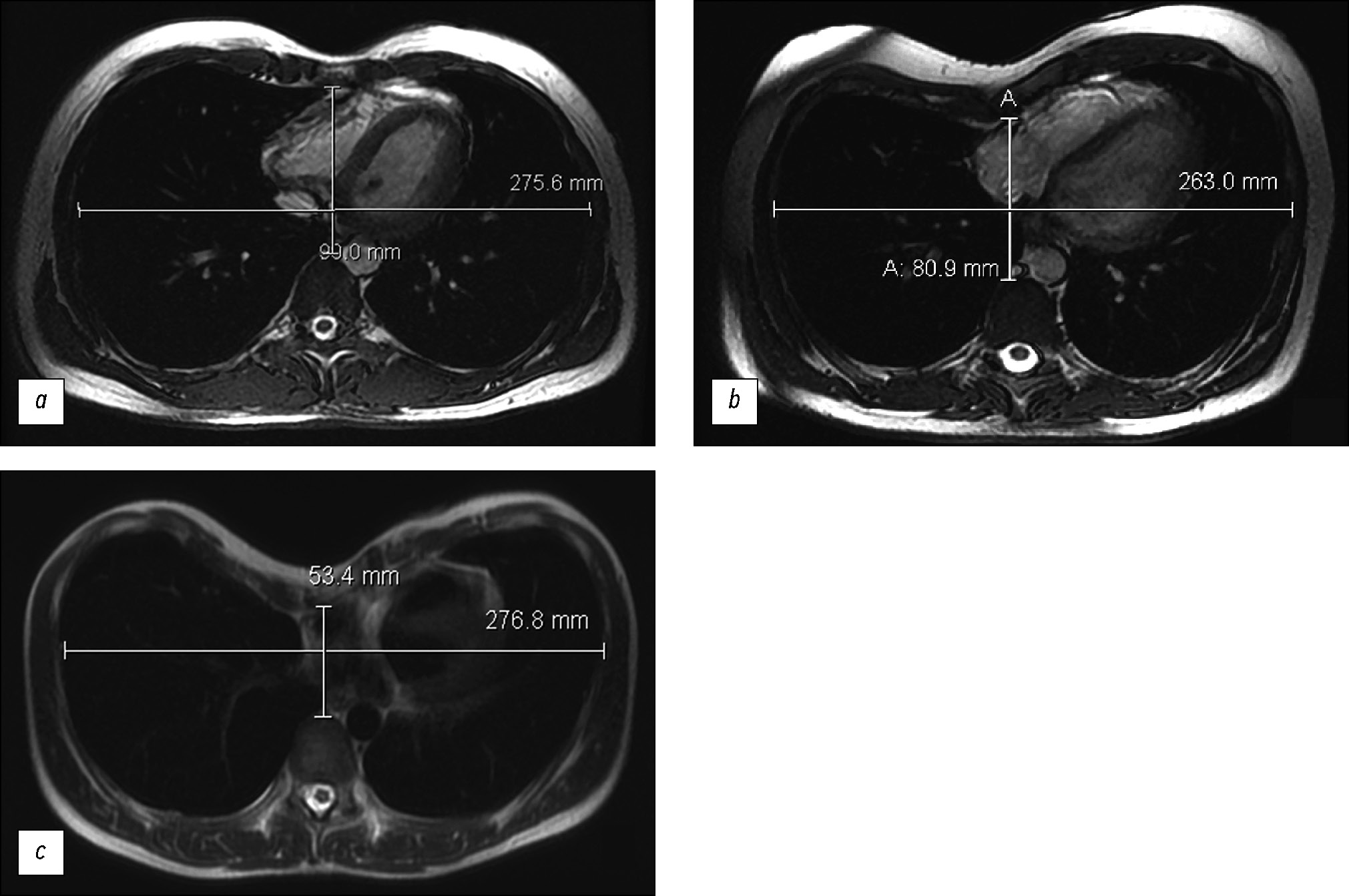
心脏磁共振成像是在 General Electric Optima MR450w GEM 1.5 特斯拉设备上进行的,使用的是 2D-FIESTA-C 脉冲序列,同时进行了伴有左心室和右心室功能评估的心电图同步。获得了对漏斗胸畸形患者进行进一步手术治疗所需的参数:Haller 指数、矫正指数、胸骨旋转角度。
对获得的数据进行统计处理,寻找 Haller 指数、矫正指数、胸骨旋转角度和右心室射血分数之间的相关性。P<0.05的值被认为是统计学意义的边界。
结果。在 92% 的患者中发现了中度和重度漏斗胸畸形。在寻找 Haller指数值与右心室射血分数之间的相关性时,未发现有统计学意义的 Pearson 相关性(吸气射血分数值的相关性为 P=0.777,呼气射血分数值的相关性为 P=0.798)。右心室射血分数的平均值为 46%。在统计分析中,随着 Haller 指数(胸腔器官畸形程度的增加)的增加,矫正指数也在增加(P<0.05)。44.7% 的患者的胸骨旋转角度值需要修改手术干预(超过15°)。
结论。磁共振成像是一种对漏斗胸畸形有高度参考价值的诊断方法:无需放射线照射,还能对病理变化进行详细的术前评估。
数据显示了,Haller 指数与矫正指数值之间存在相关性(P<0.05)。此外,磁共振成像数据显示了,右心室射血分数有所下降。
作者简介
Gulishe S. Muzafarova
Moscow Regional Research and Clinical Institute
Email: gms0495@mail.ru
ORCID iD: 0000-0003-0940-3247
SPIN 代码: 2950-5431
俄罗斯联邦, Moscow
Marina V. Vishnyakova
Moscow Regional Research and Clinical Institute
编辑信件的主要联系方式.
Email: cherridra@mail.ru
ORCID iD: 0000-0003-3838-636X
SPIN 代码: 1137-2991
MD, Dr. Sci. (Medicine)
俄罗斯联邦, MoscowAlexander S. Abramenko
Moscow Regional Research and Clinical Institute
Email: a.s.abramenko@gmail.com
ORCID iD: 0000-0002-6286-2162
SPIN 代码: 9743-3001
俄罗斯联邦, Moscow
Vladimir A. Kuzmichev
Moscow Regional Research and Clinical Institute
Email: vakuzmichev@gmail.ru
ORCID iD: 0000-0001-6493-8012
SPIN 代码: 8345-5298
MD, Cand. Sci. (Medicine), Professor
俄罗斯联邦, MoscowVladimir V. Gatsutsyn
Moscow Regional Research and Clinical Institute
Email: vg86@list.ru
ORCID iD: 0000-0002-2364-5325
SPIN 代码: 1431-4417
俄罗斯联邦, Moscow
参考
- Pechetov AA, Esakov JuS, Gubajdullina GF, Makov MA, Hlan’ TN. Differetial approach for chest wall reconstruction for pectus excavatum for adult. N.I. Pirogov Journal of Surgery. 2017;(7):24–29. doi: 10.17116/hirurgia2017724-29
- Fokin АА, Steuerwald NM, Ahrens WA, Allen KE. Anatomical, histologic, and genetic characteristics of congenital chest wall deformities. Seminars in Thoracic and Cardiovascular Surgery. 2009;21(1):44–57. doi: 10.1053/j.semtcvs.2009.03.001
- Scalise PN, Demehri FR. The management of pectus excavatum in pediatric patients: a narrative review. Transl Pediatr. 2023;12(2):208–220. doi: 10.21037/tp-22-361
- Trò R, Martini S, Stagnaro N, et al. A new tool for assessing Pectus Excavatum by a semi-automatic image processing pipeline calculating the classical severity indexes and a new marker: the Volumetric Correction Index. BMC Med Imaging. 2022. doi: 10.1186/s12880-022-00754-0
- Andreyev PS, Skvortsov AP, Tsoy IV, et al. Treatment of funnel breast in children and adolescents. Practical medicine. 2021;19(4):138–141. doi: 10.32000/2072-1757-2021-4-138-141
- Andreev PS, Skvortsov AР, Khabibyanov RYa, Maleev MV. Our experience in surgical treatment of penetral chest deformation. Annali d’Italia. 2023;(41):53–57. doi: 10.5281/zenodo.7774296
- Haller JA Jr, Kramer SS, Lietman SA, et al. Use of CT scans in selection of patients for pectus excavatum surgery: a preliminary report. J Pediatr Surg. 1987;22(10):904–906. doi: 10.1016/s0022-3468(87)80585-7
- Sidden CR, Katz ME, Swoveland BC, Nuss D. Radiologic considerations in patients undergoing the Nuss procedure for correction of pectus excavatum. Pediatric Radiology. 2001;31(6):429–434. doi: 10.1007/s002470100455
- St. Peter SD, Juang D, Garey CL, et al. A novel measure for pectus excavatum: the correction index. Journal of Pediatric Surgery. 2011;46(12):2270–2273. doi: 10.1016/j.jpedsurg.2011.09.009
- Tauchi R, Suzuki Y, Tsuji T, et al. Clinical Characteristics and Thoracic factors in patients with Idiopathic and Syndromic Scoliosis Associated with Pectus Excavatum. Spine Surg Relat Res. 2018;2(1):37–41. doi: 10.22603/ssrr.2017-0027
- Shamsiev AM, Shamsiev ZhA, Turaev JuA, Mutalibov AI, Burgutov MZh. The role of functional studies of the cardiorespiratory system with funnel chest deformity. Journal Problems of Biology and Medicine. 2017;1(93):9–14.
- Peng R, Mardakhaev E, Shmukler A, Levsky JM, Haramati LB. Meeting ACR Dose Guidelines for CT Lung Cancer Screening in an Overweight and Obese Population. Acad Radiol. 2021;28(3):381–386. doi: 10.1016/j.acra.2020.02.009
- Mortellaro VE, Iqbal CW, Fike FB, et al. The predictive value of Haller index in patients undergoing pectus bar repair for pectus excavatum. J Surg Res. 2011;170(1):104–106. doi: 10.1016/j.jss.2011.02.014
- Karakılıç A, Karaçam V, Ersöz H, et al. Determination of severity of deformity with rib length to costal cartilage length ratio in thorax deformities. Turk Gogus Kalp Damar Cerrahisi Derg. 2018;26(2):279–285. doi: 10.5606/tgkdc.dergisi.2018.15009
- Poston PM, Patel SS, Rajput M, et al. The correction index: setting the standard for recommending operative repair of pectus excavatum. Ann Thorac Surg. 2014;97(4):1176–1180. doi: 10.1016/j.athoracsur.2013.12.050
- Marcovici PA, LoSasso BE, Kruk P, Dwek JR. MRI for the evaluation of pectus excavatum. Pediatric Radiology. 2011;41:757–758. doi: 10.1007/s00247-011-2031-5
- Lollert A, Funk J, Tietze N, et al. Morphologic assessment of thoracic deformities for the preoperative evaluation of pectus excavatum by magnetic resonance imaging. European Radiology. 2015;25:785–791. doi: 10.1007/s00330-014-3450-0
- Dore M, Triana JP, Bret M, et al. Advantages of Cardiac Magnetic Resonance Imaging for Severe Pectus Excavatum Assessment in Children. Eur J Pediatr Surg. 2018;28(1):34–38. doi: 10.1055/s-0037-1604427
- Saleh RS, Finn JP, Fenchel M, et al. Cardiovascular magnetic resonance in patients with pectus excavatum compared with normal controls. J Cardiovasc Magn Reson. 2010;12(1). doi: 10.1186/1532-429X-12-73
- Stagnaro N, Trocchio G, Torre M, et al. Cardiovascular MRI assessment of pectus excavatum in pediatric patients and postoperative simulation using vacuum bell. J Pediatr Surg. 2021;56(9):1600–1605. doi: 10.1016/j.jpedsurg.2020.11.017
补充文件


