

")


Imaging techniques in diagnosing acute pulmonary thromboembolism
- Authors: Oganesyan A.A.1, Sinitsyn V.E.2, Mershina E.A.2, Pershina E.S.1
-
Affiliations:
- Pirogov Municipal Clinical Hospital No. 1
- Lomonosov Moscow State University
- Issue: Vol 6, No 1 (2025)
- Pages: 130-142
- Section: Reviews
- URL: https://ogarev-online.ru/DD/article/view/310057
- DOI: https://doi.org/10.17816/DD634639
- ID: 310057
Cite item

Abstract
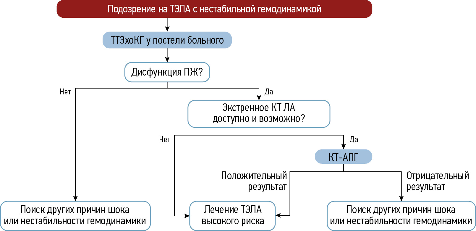
Pulmonary thromboembolism is the occlusion of the pulmonary arteries by thrombi of any origin, which commonly originates in the large veins of the legs and pelvis. This article provides an overview of existing imaging techniques used in diagnosing this pathology. A review of scientific studies by Russian and international authors is provided. Moreover, the article discusses diagnostic algorithms and the characteristics and challenges of risk stratification in patients with suspected acute pulmonary thromboembolism. The key imaging aspects for this pathology and criteria for assessing its severity are highlighted. The contribution of relatively new perfusion tomography methods, such as dual-energy and subtraction computed tomography pulmonary angiography, and magnetic resonance pulmonary angiography is demonstrated. Despite the presence of established methods for diagnosing acute pulmonary embolism, there is growing interest in additional and alternative imaging techniques, which have been more integrated into routine clinical practice. Special attention is given to subtraction computed tomography pulmonary angiography, which has the ability to generate iodine maps for indirect perfusion assessment, and its application in clinical practice. The feasibility of using various imaging techniques in diagnosing acute pulmonary thromboembolism is discussed, highlighting their advantages and prospects in emergency medical care.
Full Text
##article.viewOnOriginalSite##About the authors
Anait A. Oganesyan
Pirogov Municipal Clinical Hospital No. 1
Author for correspondence.
Email: talilen@mail.ru
ORCID iD: 0000-0003-1896-023X
SPIN-code: 6531-2957
Russian Federation, Moscow
Valentin E. Sinitsyn
Lomonosov Moscow State University
Email: vsini@mail.ru
ORCID iD: 0000-0002-5649-2193
SPIN-code: 8449-6590
MD, Dr. Sci. (Medicine), Professor
Russian Federation, MoscowElena A. Mershina
Lomonosov Moscow State University
Email: Elena_Mershina@mail.ru
ORCID iD: 0000-0002-1266-4926
SPIN-code: 6897-9641
MD, Cand. Sci. (Medicine), Assistant Professor
Russian Federation, MoscowEkaterina S. Pershina
Pirogov Municipal Clinical Hospital No. 1
Email: pershina@mail.ru
ORCID iD: 0000-0002-3952-6865
SPIN-code: 7311-9276
MD, Cand. Sci. (Medicine)
Russian Federation, MoscowReferences
- Vertkin AL, Gritsanchuk AM. Tromboembolism: an epidemic that everyone is silent about. The Russian Archives of Internal Medicine. 2014;(1):33–39. (In Russ.) doi: 10.20514/2226-6704-2014-0-1-33-39 EDN: TBCLKL
- Goldhaber SZ, Visani L, De Rosa M. Acute pulmonary embolism: clinical outcomes in the International Cooperative Pulmonary Embolism Registry (ICOPER). The Lancet. 1999;353(9162):1386–1389. doi: 10.1016/S0140-6736(98)07534-5 EDN: DAROEL
- Kucher N, Rossi E, De Rosa M, Goldhaber SZ. Massive pulmonary embolism. Circulation. 2006;113(4):577–582. doi: 10.1161/CIRCULATIONAHA.105.592592
- Bajc M, Schümichen C, Grüning T, et al. EANM guideline for ventilation/perfusion single-photon emission computed tomography (SPECT) for diagnosis of pulmonary embolism and beyond. European Journal of Nuclear Medicine and Molecular Imaging. 2019;46(12):2429–2451. doi: 10.1007/s00259-019-04450-0 EDN: QGKIJW
- Mukhametshina GA, Amirov NB, Frolova EB, et al. The qustion of pulmonary artery tromboembolism. The Bulletin of Contemporary Clinical Medicine. 2013;6(4):67–73. EDN: RCKYBF
- Zhuravkov YuL, Koroleva AA. Modern principles of diagnosis and treatment of the acute pulmonary thromboembolism. Military Medicine. 2014;3(32):112–116. EDN: SLQPTP
- Sukhova MB, Trofimova TN. Modern aspects of MSCT diagnostics of acute massive pulmonary embolism. Diagnostic radiology and radiotherapy. 2021;12(4):7–14. doi: 10.22328/2079-5343-2021-12-4-7-14 EDN: OHOTRM
- Bokeriya LA, Zatevakhin II, Kirienko AI, et al. Russian clinical guidelines for the diagnosis, treatment and prevention of venous thromboembolic complications (VTEC). Flebologiya. 2015;9(4-2):1–52 (In Russ.) EDN: XIOPYZ
- Tyurin IE. Pulmonary embolism: possibilities of radiological diagnostics. Atmosfera. Pul'monologiya i allergologiya. 2005;(4):20–24 (In Russ.) EDN: OOPMTF
- Heit JA. Venous thromboembolism epidemiology: implications for prevention and management. Seminars in thrombosis and hemostasis. 2002;28 Suppl. 2:3–13. doi: 10.1055/s-2002-32312
- Sweet PH 3rd, Armstrong T, Chen J, et al. Fatal pulmonary embolism update: 10 years of autopsy experience at an academic medical center. JRSM Short Reports. 2013;4(9):2042533313489824. doi: 10.1177/2042533313489824
- Konstantinides SV, Meyer G, Becattini C, et al. 2019 ESC Guidelines for the diagnosis and management of acute pulmonaryembolism developed in collaboration with the European Respiratory Society (ERS). Russian Journal of Cardiology. 2020;25(8):180–239. doi: 10.15829/1560-4071-2020-3848 EDN: NXTZZJ
- Ceriani E, Combescure C, Le Gal G, et al. Clinical prediction rules for pulmonary embolism: a systematic review and meta-analysis. Journal of Thrombosis and Haemostasis. 2010;8(5):957–970. doi: 10.1111/j.1538-7836.2010.03801.x
- Quezada CA, Bikdeli B, Barrios D, et al. Meta-analysis of prevalence and short-term prognosis of hemodynamically unstable patients with symptomatic acute pulmonary embolism. The American Journal of Cardiology. 2019;123(4):684–689. doi: 10.1016/j.amjcard.2018.11.009
- Singh R, Nie RZ, Homayounieh F, et al. Quantitative lobar pulmonary perfusion assessment on dual-energy CT pulmonary angiography: applications in pulmonary embolism. European Radiology. 2020;30(5):2535–2542. doi: 10.1007/s00330-019-06607-9 EDN: QVXDOT
- Tafur AJ, Shamoun FE, Patel SI, et al. Catheter-directed treatment of pulmonary embolism: a systematic review and meta-analysis of modern literature. Clinical and Applied Thrombosis/Hemostasis. 2016;23(7):821–829. doi: 10.1177/1076029616661414
- Kröger JR, Hickethier T, Pahn G, et al. Influence of spectral detector CT based monoenergetic images on the computer-aided detection of pulmonary artery embolism. European Journal of Radiology. 2017;95:242–248. doi: 10.1016/j.ejrad.2017.08.034
- Qanadli S, El Hajjam M, Vieillard-Baron A, et al. New CT index to quantify arterial obstruction in pulmonary embolism. American Journal of Roentgenology. 2001;176(6):1415–1420. doi: 10.2214/ajr.176.6.1761415
- Frank Peacock W, Coleman CI, Diercks DB, et al. Emergency department discharge of pulmonary embolus patients. Academic Emergency Medicine. 2018;25(9):995–1003. doi: 10.1111/acem.13451
- Torbicki A, Perrier A, Konstantinides S, et al. Guidelines on the diagnosis and management of acute pulmonary embolism: the task force for the diagnosis and management of acute pulmonary embolism of the European Society of Cardiology (ESC). European Heart Journal. 2008;29(18):2276–2315. doi: 10.1093/eurheartj/ehn310
- Giordano J, Khung S, Duhamel A, et al. Lung perfusion characteristics in pulmonary arterial hypertension (PAH) and peripheral forms of chronic thromboembolic pulmonary hypertension (pCTEPH): dual-energy CT experience in 31 patients. European Radiology. 2017;27(4):1631–1639. doi: 10.1007/s00330-016-4500-6 EDN: WXUBJW
- Pietersen PI, Goyard C, Gill T, et al. The CT revolution: the role of PIOPED II in establishing CT pulmonary angiography as the reference standard for pulmonary embolism diagnosis. Breathe (Sheff). 2024; 20(1):230228. doi: 10.1183/20734735.0228-2023 EDN: LNAYLM
- Viteri-Ramírez G, García-Lallana A, Simón-Yarza I, et al. Low radiation and low-contrast dose pulmonary CT angiography: comparison of 80 kVp/60 ml and 100 kVp/80 ml protocols. Clinical Radiology. 2012;67(9):833–839. doi: 10.1016/j.crad.2011.11.016
- Gietema HA, Walraven KHM, Posthuma R, et al. Dual-energy computed tomography compared to lung perfusion scintigraphy to assess pulmonary perfusion in patients screened for endoscopic lung volume reduction. Respiration. 2021;100(12):1186–1195. doi: 10.1159/000517598 EDN: HCPPYM
- Im DJ, Hur J, Han K, et al. Prognostic value of dual-energy CT-based iodine quantification versus conventional CT in acute pulmonary embolism: a propensity-match analysis. Korean Journal of Radiology. 2020;21(9):1095. doi: 10.3348/kjr.2019.0645 EDN: ZLWQQL
- Zhang LJ, Zhou CHSH, Schoepf UJ, et al. Dual-energy CT lung ventilation/perfusion imaging for diagnosing pulmonary embolism. European Radiology. 2013;23(10):2666–2675. doi: 10.1007/s00330-013-2907-x EDN: HTDZSY
- Yang GF, Yang X, Zhang LJ, et al. Pulmonary enhancement imaging with dual energy CT for the detection of pulmonary embolism in a rabbit model. Academic Radiology. 2011;18(5):605–614. doi: 10.1016/j.acra.2010.12.012
- Nikolaou K, Tiem S, Sommer W, et al. Diagnosing pulmonary embolism: new computed tomography applications. Journal of Thoracic Imaging. 2010;25(2):151–160. doi: 10.1097/RTI.0b013e3181d9ca1d
- Zhang LJ, Wang ZJ, Zhou CS, et al. Evaluation of pulmonary embolism in pediatric patients with nephrotic syndrome with dual energy CT pulmonary angiography. Academic Radiology. 2012;19(3):341–348. doi: 10.1016/j.acra.2011.11.002
- Ruggiero A, Screaton NJ. Imaging of acute and chronic thromboembolic disease: state of the art. Clinical Radiology. 2017;72(5):375–388. doi: 10.1016/j.crad.2017.02.011
- Otrakji A, Digumarthy SR, Lo Gullo R, et al. Dual-energy CT: spectrum of thoracic abnormalities. RadioGraphics. 2016;36(1):38–52. doi: 10.1148/rg.2016150081
- Rassouli N, Etesami M, Dhanantwari A, Rajiah P. Detector-based spectral CT with a novel dual-layer technology: principles and applications. Insights into Imaging. 2017;8(6):589–598. doi: 10.1007/s13244-017-0571-4 EDN: RIGZBG
- Ermolaev VL, Stolin AV, Shurygina EP, et al. Capabilities of traditional integrated approach to diagnosis and management of acute pulmonary embolism. Ural Medical Journal. 2010;7(72):58–62. EDN: MWJYVB
- Zabavskaya OA, Sharifullin FA, Kokov LS. Capabilities of multispiral computer tomography in differential diagnosis of pulmonary embolism. In: Proceedings of the scientific and practical conference «New technologies in emergency and urgent medical care». Suzdal, 2016 Apr 21–22. Moscow: N.V. Sklifosovsky Research Institute of Emergency Care. P. 131–132. (In Russ.)
- Perrier A, Howarth N, Didier D, et al. Performance of helical computed tomography in unselected outpatients with suspected pulmonary embolism. Annals of Internal Medicine. 2001;135(2):88. doi: 10.7326/0003-4819-135-2-200107170-00008
- Van Strijen MJ, De Monye W, Kieft GJ, et al. Accuracy of single-detector spiral CT in the diagnosis of pulmonary embolism: a prospective multicenter cohort study of consecutive patients with abnormal perfusion scintigraphy. Journal of Thrombosis and Haemostasis. 2005;3(1):17–25. Corrected and republished from: Journal of Thrombosis and Haemostasis. 2005;3(3):622. doi: 10.1111/j.1538-7836.2004.01064.x
- Mullins MD, Becker DM, Hagspiel KD, Philbrick JT. The role of spiral volumetric computed tomography in the diagnosis of pulmonary embolism. Archives of Internal Medicine. 2000;160(3):293–298. doi: 10.1001/archinte.160.3.293
- Prokop M, Galanski M, editors. Spiral and multislice computer tomography of the body. New York: Thieme; 2003.
- Kelly AM. Imaging in thromboembolic disease. Imaging Med. 2011;3(1):31–50.
- Gottschalk A, Sostman HD, Coleman RE, et al. Ventilation-perfusion scintigraphy in the PIOPED study. Part II. Evaluation of the scintigraphic criteria and interpretations. J Nucl Med. 1993;34(7):1119–1126.
- Waterstram-Rich KM, Gilmore D. Respiratory system. In: Gilmore D, Waterstram-Rich KM. Nuclear Medicine and PET/CT: technology and techniques. 8th ed. St. Louis, MO: Mosby Elsevier; 2016. P. 475–487.
- Chan K, Ioannidis S, Coghlan JG, et al. Pulmonary arterial hypertension with abnormal V/Q single-photon emission computed tomography. JACC: Cardiovascular Imaging. 2018;11(10):1487–1493. doi: 10.1016/j.jcmg.2017.07.026 EDN: RDHSTD
- Yu L, Leng S, McCollough CH. Dual-energy CT–based monochromatic imaging. American Journal of Roentgenology. 2012;199(suppl. 5):S9–S15. doi: 10.2214/AJR.12.9121
- Lenga L, Trapp F, Albrecht MH, et al. Single- and dual-energy CT pulmonary angiography using second- and third-generation dual-source CT systems: comparison of radiation dose and image quality. European Radiology. 2019;29(9):4603–4612. doi: 10.1007/s00330-018-5982-1 EDN: YGLIHW
- Weidman EK, Plodkowski AJ, Halpenny DF, et al. Dual-energy CT angiography for detection of pulmonary emboli: incremental benefit of iodine maps. Radiology. 2018;289(2):546–553. doi: 10.1148/radiol.2018180594
- Masy M, Giordano J, Remy J, et al. Dual-energy CT (DECT) lung perfusion in pulmonary hypertension: concordance rate with V/Q scintigraphy in diagnosing chronic thromboembolic pulmonary hypertension (CTEPH). European Radiology. 2018;28(12):5100–5110. doi: 10.1007/s00330-018-5467-2 EDN: HBNJYG
- Takx RAP, Henzler T, Schoepf UJ, et al. Predictive value of perfusion defects on dual energy CTA in the absence of thromboembolic clots. Journal of Cardiovascular Computed Tomography. 2017;11(3):183–187. doi: 10.1016/j.jcct.2017.04.005
- Mershina EA, Sinitsyn VE, Plotnikova ML,et al. Use of dual-energy computed tomographic angiopulmonography in patients with chronic thromboembolic pulmonary hypertension before and after pulmonary artery thromboendarterectomy. Journal of Radiology and Nuclear Medicine. 2013;2:27–31. EDN: RGQWTT
- Ohno Y, Ozawa Y, Nagata H, et al. Area-detector computed tomography for pulmonary functional imaging. Diagnostics. 2023;13(15):2518. doi: 10.3390/diagnostics13152518 EDN: LXLBNJ
- Hong YJ, Shim J, Im DJ, et al. Dual-energy CT for pulmonary embolism: current and evolving clinical applications. Korean Journal of Radiology. 2021;22(9):1555–1568. doi: 10.3348/KJR.2020.1512 EDN: EFSHLY
- Tamura M, Yamada Y, Kawakami T, et al. Diagnostic accuracy of lung subtraction iodine mapping CT for the evaluation of pulmonary perfusion in patients with chronic thromboembolic pulmonary hypertension: correlation with perfusion SPECT/CT. International Journal of Cardiology. 2017;243:538–543. doi: 10.1016/j.ijcard.2017.05.006
- Shahin Y, Johns C, Karunasaagarar K, et al. IodiNe subtraction mapping in the diagnosis of pulmonary chronIc thRomboEmbolic disease (INSPIRE): rationale and methodology of a cross-sectional observational diagnostic study. Contemporary Clinical Trials Communications. 2019;15:100417. doi: 10.1016/j.conctc.2019.100417
- Tsuchiya N, van Beek EJR, Ohno Y, et al. Magnetic resonance angiography for the primary diagnosis of pulmonary embolism: a review from the international workshop for pulmonary functional imaging. World Journal of Radiology. 2018;10(6):52–64. doi: 10.4329/wjr.v10.i6.52
- Fu Q, Cheng Q, Kong X, et al. Diagnostic accuracy of true fast imaging with steady-state precession, MR pulmonary angiography and volume-interpolated body examination for pulmonary embolism compared with CT pulmonary angiography. Experimental and Therapeutic Medicine. 2020;21(1):42. doi: 10.3892/etm.2020.9474 EDN: VLQMDT
Supplementary files


