

")


利用人工智能检测肺癌新病例:COVID-19大流行2年后计算机断层扫描结果回顾性分析的临床和经济评估
- 作者: Zukov R.A.1,2, Safontsev I.P.1,2, Klimenok M.P.2, Zabrodskaya T.E.2, Merkulova N.A.2, Chernina V.Y.3, Belyaev M.G.3, Goncharov M.Y.3,4,5, Omelyanovskiy V.V.6,7,8, Ulianova K.A.9, Soboleva E.A.3,5, Blokhina M.E.10, Nalivkina E.A.10, Gombolevskiy V.A.3,4,11,12
-
隶属关系:
- Professor V.F. Voino-Yasenetsky Krasnoyarsk State Medical University
- Krasnoyarsk Regional Clinical Oncological Dispensary named after A.I. Kryzhanovskogo
- IRA Labs
- Artificial Intelligence Research Institute AIRI
- Skolkovo Institute of Science and Technology
- Center for Expertise and Quality Control of Medical Care
- Russian Medical Academy of Continuous Professional Education
- Financial Research Institute
- Ministry of Health of the Russian Federation
- AstraZeneca Pharmaceuticals LLC
- World-Class Research Center «Digital biodesign and personalized healthcare»
- Sechenov First Moscow State Medical University
- 期: 卷 5, 编号 4 (2024)
- 页面: 725-739
- 栏目: 原创性科研成果
- URL: https://ogarev-online.ru/DD/article/view/309832
- DOI: https://doi.org/10.17816/DD630885
- ID: 309832
如何引用文章

详细
论证。胸腔器官计算机断层扫描是COVID-19感染引起的肺组织变化的主要诊断方法。因此,自2020年以来,这项研究在克拉斯诺亚尔斯克边疆区的应用频率有所增加。然而,肺癌的发病率却下降了5.2%。这种情况引起了人们对肺癌漏检特征性放射学变化的担忧,并促使人们寻找新的诊断技术,包括用于数据分析的人工智能(AI)。
目的 — 评估使用人工智能算法从COVID-19大流行期间获得的胸腔器官计算机断层扫描数据中,搜索肺结节发现肺癌的可行性。
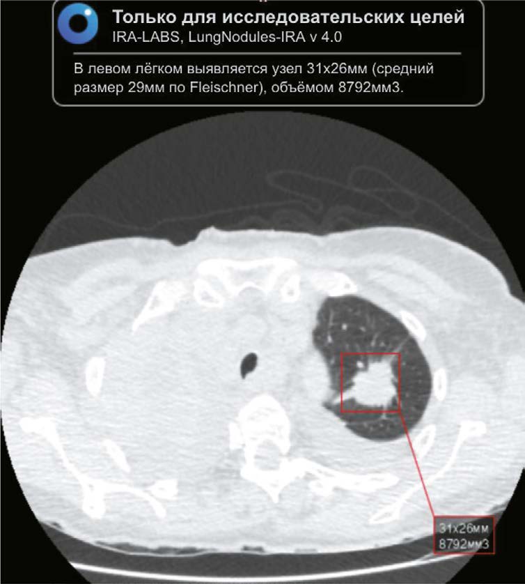
材料和方法。这项回顾性研究,包括从2020年1月11日至 2021年2月28日期间,克拉斯诺亚尔斯克地区 从PACS档案中诊断为COVID-19的患者的胸腔器官计算机断层扫描结果。进行胸腔器官计算机断层扫描与应用人工智能算法之间的时间间隔从两年零一个月到两年零五个月。使用了Chest-IRA AI算法。AI检测到体积大于100mm3的肺部结节。放射科医生根据肺癌的概率将结果分为三组。使用该算法的经济效益评估,考虑到了工资成本和早期治疗肺癌所节省的费用,这些也会影响地区生产总值。
结果。在10500个计算机断层扫描结果中,人工智能算法检查出484例结节性肿块。确定了192名患肺癌高概率的患者,103名无体征,60名体征根据不足。112名肺癌高概率和中概率的患者没有就医。 通过使用人工智能,100例(28.2%)经组织学验证的肺癌患者得到了确诊,其中35%的患者处于I-II期。
使用人工智能代替放射科医生可以节省25个月零4 天的工作时间,也就是243万卢布。每进行10500次计算机断层扫描,因早期发现肺癌而节省的预算预计从1060万卢布到1250万卢布。五年的总经济效益为2.594亿卢布至3.051亿卢布。
结论。使用人工智能分析胸腔器官计算机断层扫描结果显示肺结节检测的高效率,包括在COVID-19的背景下,这证实其用于早期发现那些可能被遗漏的随机肺结节的前景。
作者简介
Ruslan A. Zukov
Professor V.F. Voino-Yasenetsky Krasnoyarsk State Medical University; Krasnoyarsk Regional Clinical Oncological Dispensary named after A.I. Kryzhanovskogo
编辑信件的主要联系方式.
Email: zukov_rus@mail.ru
ORCID iD: 0000-0002-7210-3020
SPIN 代码: 3632-8415
MD, Dr. Sci. (Medicine), Professor
俄罗斯联邦, Krasnoyarsk; KrasnoyarskIvan P. Safontsev
Professor V.F. Voino-Yasenetsky Krasnoyarsk State Medical University; Krasnoyarsk Regional Clinical Oncological Dispensary named after A.I. Kryzhanovskogo
Email: sip@onkolog24.ru
ORCID iD: 0000-0002-8177-6788
SPIN 代码: 1548-5565
MD, Cand. Sci. (Medicine), Assoc. Prof., Depart. of Oncology and Radiation Therapy with a Postgraduate Course, Deputy Head Physician
俄罗斯联邦, Krasnoyarsk; KrasnoyarskMarina P. Klimenok
Krasnoyarsk Regional Clinical Oncological Dispensary named after A.I. Kryzhanovskogo
Email: klimenokmp@onkolog24.ru
ORCID iD: 0009-0001-7849-0770
SPIN 代码: 7179-8793
MD
俄罗斯联邦, KrasnoyarskTatyana E. Zabrodskaya
Krasnoyarsk Regional Clinical Oncological Dispensary named after A.I. Kryzhanovskogo
Email: ZabrodskayaTE@onkolog24.ru
ORCID iD: 0000-0003-4987-5222
SPIN 代码: 8365-3582
MD
俄罗斯联邦, KrasnoyarskNatalya A. Merkulova
Krasnoyarsk Regional Clinical Oncological Dispensary named after A.I. Kryzhanovskogo
Email: MerkulovaNA@onkolog24.ru
ORCID iD: 0009-0006-9254-1331
MD
俄罗斯联邦, KrasnoyarskValeria Yu. Chernina
IRA Labs
Email: v.chernina@ira-labs.com
ORCID iD: 0000-0002-0302-293X
SPIN 代码: 8896-8051
MD
俄罗斯联邦, MoscowMikhail G. Belyaev
IRA Labs
Email: belyaevmichel@gmail.com
ORCID iD: 0000-0001-9906-6453
SPIN 代码: 2406-1772
Cand. Sci. (Physics and Mathematics)
俄罗斯联邦, MoscowMikhail Yu. Goncharov
IRA Labs; Artificial Intelligence Research Institute AIRI; Skolkovo Institute of Science and Technology
Email: mig0nch@yandex.ru
ORCID iD: 0009-0009-8417-0878
俄罗斯联邦, Moscow; Moscow; Moscow
Vitaly V. Omelyanovskiy
Center for Expertise and Quality Control of Medical Care; Russian Medical Academy of Continuous Professional Education; Financial Research Institute
Email: vvo@rosmedex.ru
ORCID iD: 0000-0003-1581-0703
SPIN 代码: 1776-4270
MD, Dr. Sci. (Medicine), Professor
俄罗斯联邦, Moscow; Moscow; MoscowKsenia A. Ulianova
Ministry of Health of the Russian Federation
Email: UlyanovaKA@minzdrav.gov.ru
ORCID iD: 0000-0002-3462-0123
SPIN 代码: 6491-6072
俄罗斯联邦, Moscow
Evgenia A. Soboleva
IRA Labs; Skolkovo Institute of Science and Technology
Email: e.soboleva@ira-labs.com
ORCID iD: 0009-0009-4037-6911
俄罗斯联邦, Moscow; Moscow
Maria E. Blokhina
AstraZeneca Pharmaceuticals LLC
Email: mariya.blokhina@astrazeneca.com
ORCID iD: 0009-0002-9008-9485
MD
俄罗斯联邦, MoscowElena A. Nalivkina
AstraZeneca Pharmaceuticals LLC
Email: elena.nalivkina@astrazeneca.com
ORCID iD: 0009-0003-5412-9643
俄罗斯联邦, Moscow
Victor A. Gombolevskiy
IRA Labs; Artificial Intelligence Research Institute AIRI; World-Class Research Center «Digital biodesign and personalized healthcare»; Sechenov First Moscow State Medical University
Email: gombolevskii@gmail.com
ORCID iD: 0000-0003-1816-1315
SPIN 代码: 6810-3279
MD, Cand. Sci. (Medicine)
俄罗斯联邦, Moscow; Moscow; Moscow; Moscow参考
- Siegel RL, Miller KD, Jemal A. Cancer statistics, 2020. CA Cancer J Clin. 2020;70(1):7–30. doi: 10.3322/caac.21590
- Kaprin AD, Starinsky VV, Shakhzadova AO, editors. Malignant neoplasms in Russia in 2021 (morbidity and mortality). Moscow: P. Herzen MORI – the branch of the FSBI NMRRC of the Ministry of Health of the Russian Federation, 2022. (In Russ).
- Сhest-IRA [Internet]; 2020. [cited 2024 May 16]. Available from: https://mosmed.ai/service_catalog/chestira/
- Morozov SP, Vladzimirsky AV, Klyashtorny VG, et al. Clinical trials of software based on intelligent technologies (Radiology). Series «Best Practices of Radiology and Instrumental Diagnostics». N 56. Moscow: SBHI «SPCC for DTT of MHD», 2019. (In Russ).
- Armato SG 3rd, McLennan G, Bidaut L, et al. The lung image database consortium (LIDC) and image database resource initiative (IDRI): a completed reference database of lung nodules on CT scans. Med Phys. 2011;38(2):915–931. doi: 10.1118/1.3528204
- Goncharov M, Pisov M, Shevtsov A, et al. CT-Based COVID-19 triage: dep multitask learning improves joint identification and severity quantification. Med Image Anal. 2021;71:102054. doi: 10.1016/j.media.2021.102054
- MacMahon H, Naidich DP, Goo JM, et al. Guidelines for management of incidental pulmonary nodules detected on CT images: from the Fleischner Society 2017. Radiology. 2017;284(1):228–243. doi: 10.1148/radiol.2017161659
- Rate agreement of the compulsory medical system of Krasnoyarsk Territory. In: territorial fund of compulsory medical insurance; 2012– [cited 2024 May 16]. Available from: https://www.krasmed.ru/content/18137/page.html
- Kaprin AD, Starinsky VV, Shakhzadova AO. State of oncological care for the Russian population in 2021. Moscow: P. Herzen MORI — the branch of the FSBI NMRRC of the Ministry of Health of the Russian Federation, 2022. (In Russ).
- National Lung Screening Trial Research Team. Reduced lung cancer mortality with low dose computed tomographic screening. N Engl J Med. 2011;365(5):395–409. doi: 10.1056/NEJMoa1102873
- Gusamova NV, Komleva MI, Saphontsev IP, et al. Lung cancer screening by LDCT. Results for 2015–2017 years RSBHCI «Krasnoyarsk Regional Clinical Oncological Dispensary named after A.I. Kryzhanovsky». In: Modern achievements of oncology in clinical practice. Proceedings of the All Russian scientific and practical conference. Krasnoyarsk, 2018. P. 54–57. (In Russ).
- Ardila D, Kiraly AP, Bharadwaj S, et al. End to end lung cancer screening with three dimensional deep learning on low dose chest computed tomography. Nat Med. 2019;25(6):954–961. doi: 10.1038/s41591-019-0447-x
- Goncalves S, Fong PC, Blokhina M. Artificial intelligence for early diagnosis of lung cancer through incidental nodule detection in low and middle income countries acceleration during the COVID-19 pandemic but here to stay. Am J Cancer Res. 2022;12(1):1–16.
- Graf M, Makowski M, Gawlitza J, Gassert F. Cost–effectiveness of artificial intelligence support in computed tomography–based lung cancer screening. Cancers (Basel). 2022;14(7):1729. doi: 10.3390/cancers14071729
补充文件


