

")


将活检标本的组织学检查作为诊断“金标准”的局限性:一个例子
- 作者: Akhmedzyanova D.A.1, Yutsevich O.K.2, Reshetnikov R.V.1, Tashchyаn O.V.3, Pirogov S.S.2, Mazurova M.P.2, Volchenko N.N.2, Kamalov A.K.2, Shumskaya Y.F.1, Mnatsakanyan M.G.3
-
隶属关系:
- Research and Practical Clinical Center for Diagnostics and Telemedicine Technologies
- P.A. Herzen Moscow Oncology Research Institute, Branch, National Medical Research Radiological Center
- The First Sechenov Moscow State Medical University
- 期: 卷 4, 编号 4 (2023)
- 页面: 633-642
- 栏目: 临床病例及临床病例的系列
- URL: https://ogarev-online.ru/DD/article/view/262990
- DOI: https://doi.org/10.17816/DD561354
- ID: 262990
如何引用文章

详细
食管腺癌是胃肠道最常见的恶性肿瘤之一。为了在早期阶段发现这种疾病,医生采用内窥镜、形态学、免疫组化等检查方法。但是,这些方法不仅需要使用高度专业化的设备,还取决于内镜医师和病理形态学医师的专业水平。
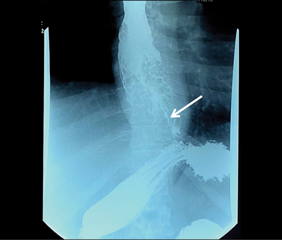
本文描述了对一名进行性吞咽困难患者的临床观察。吞咽困难是由食道肿瘤引起的。肿瘤已扩散到胃的贲门下段。肿瘤在一年内无法进行病理形态学验证。在居住地医疗机构进行的食管胃十二指肠镜检查、电子计算机断层扫描和食管双对比透视检查的数据证实了肿瘤的恶性程度。然而,大量组织学检查的结果都支持幽门腺腺瘤、顶体腺瘤或带有高度上皮发育不良病灶的肿瘤细胞腺瘤。在专业机构的条件下,通过内窥镜检查和靶向活检,才有可能证实肿瘤的恶性程度。
研究结果表明,在病理形态学检查数据相互矛盾的情况下,临床表现和仪器方法对最终诊断的重要。这再次提出活检标本的组织学检查作为诊断恶性肿瘤“金标准”的局限性问题。
作者简介
Dina A. Akhmedzyanova
Research and Practical Clinical Center for Diagnostics and Telemedicine Technologies
编辑信件的主要联系方式.
Email: AkhmedzyanovaDA@zdrav.mos.ru
ORCID iD: 0000-0001-7705-9754
SPIN 代码: 6983-5991
Scopus 作者 ID: 58104960900
俄罗斯联邦, Moscow
Olga K. Yutsevich
P.A. Herzen Moscow Oncology Research Institute, Branch, National Medical Research Radiological Center
Email: o.yutsevitch@yandex.ru
ORCID iD: 0000-0002-3860-9853
俄罗斯联邦, Moscow
Roman V. Reshetnikov
Research and Practical Clinical Center for Diagnostics and Telemedicine Technologies
Email: r.reshetnikov@npcmr.ru
ORCID iD: 0000-0002-9661-0254
SPIN 代码: 8592-0558
Cand. Sci. (Phys.-Math.)
俄罗斯联邦, MoscowOlga V. Tashchyаn
The First Sechenov Moscow State Medical University
Email: olgatash1@rambler.ru
ORCID iD: 0000-0001-6759-6820
SPIN 代码: 3658-1120
MD, Cand. Sci. (Med.)
俄罗斯联邦, MoscowSergey S. Pirogov
P.A. Herzen Moscow Oncology Research Institute, Branch, National Medical Research Radiological Center
Email: pirogov@mail.ru
ORCID iD: 0000-0002-8101-2155
SPIN 代码: 7812-5502
MD, Dr. Sci. (Med.)
俄罗斯联邦, MoscowMaria P. Mazurova
P.A. Herzen Moscow Oncology Research Institute, Branch, National Medical Research Radiological Center
Email: mnioi_morphology@mail.ru
ORCID iD: 0000-0002-4873-4455
SPIN 代码: 4455-3055
MD, Cand. Sci. (Med.)
俄罗斯联邦, MoscowNadezhda N. Volchenko
P.A. Herzen Moscow Oncology Research Institute, Branch, National Medical Research Radiological Center
Email: mnioi_morphology@mail.ru
ORCID iD: 0000-0003-0421-4172
MD, Dr. Sci. (Med.), Professor
俄罗斯联邦, MoscowAziz K. Kamalov
P.A. Herzen Moscow Oncology Research Institute, Branch, National Medical Research Radiological Center
Email: kak6768@mail.ru
ORCID iD: 0000-0001-7376-6056
SPIN 代码: 1671-1600
俄罗斯联邦, Moscow
Yuliya F. Shumskaya
Research and Practical Clinical Center for Diagnostics and Telemedicine Technologies
Email: ShumskayaYF@zdrav.mos.ru
ORCID iD: 0000-0002-8521-4045
SPIN 代码: 3164-5518
俄罗斯联邦, Moscow
Marina G. Mnatsakanyan
The First Sechenov Moscow State Medical University
Email: mnatsakanyan08@mail.ru
ORCID iD: 0000-0001-9337-7453
SPIN 代码: 2015-1822
MD, Dr. Sci. (Med.), Professor
俄罗斯联邦, Moscow参考
- Bray F, Ferlay J, Soerjomataram I, et al. Global cancer statistics 2018: GLOBOCAN estimates of incidence and mortality worldwide for 36 cancers in 185 countries. CA Cancer J Clin. 2018;68(6):394–424. doi: 10.3322/caac.21492
- McColl KEL. What is causing the rising incidence of esophageal adenocarcinoma in the West and will it also happen in the East? J Gastroenterol. 2019;54(8):669–673. doi: 10.1007/s00535-019-01593-7
- Joseph A, Raja S, Kamath S, et al. Esophageal adenocarcinoma: A dire need for early detection and treatment. Cleve Clin J Med. 2022;89(5):269–279. doi: 10.3949/ccjm.89a.21053
- Uhlenhopp DJ, Then EO, Sunkara T, Gaduputi V. Epidemiology of esophageal cancer: update in global trends, etiology and risk factors. Clin J Gastroenterol. 2020;13(6):1010–1021. doi: 10.1007/s12328-020-01237-x
- Zhang HY, Spechler SJ, Souza RF. Esophageal adenocarcinoma arising in Barrett esophagus. Cancer Lett. 2009;275(2):170–177. doi: 10.1016/j.canlet.2008.07.006
- Deng HY, Alai G, Luo J, et al. Cancerous esophageal stenosis before treatment was significantly correlated to poor prognosis of patients with esophageal cancer: a meta-analysis. J Thorac Dis. 2018;10(7):4212–4219. doi: 10.21037/jtd.2018.06.89
- Sillah K, Pritchard SA, Watkins GR, et al. The degree of circumferential tumour involvement as a prognostic factor in oesophageal cancer. Eur J Cardiothorac Surg. 2009;36(2):368–373. doi: 10.1016/j.ejcts.2008.12.052
- Deng HY, Li G, Luo J. Does oesophageal stenosis have any impact on survival of oesophageal cancer patients? Interact Cardiovasc Thorac Surg. 2018;27(3):384–386. doi: 10.1093/icvts/ivy095
- Knight WRC, McEwen R, Byrne BE, et al. Endoscopic tumour morphology impacts survival in adenocarcinoma of the oesophagus. Eur J Surg Oncol. 2020;46(12):2257–2261. doi: 10.1016/j.ejso.2020.07.003
- Morozov SP, editor. I-74 Informativeness of radial diagnostics methods in various pathological conditions of the organism. Section 2: Diagnostics of pathological conditions and diseases of the gastrointestinal tract. Moscow; 2018. (In Russ).
- Ishihara R, Goda K, Oyama T. Endoscopic diagnosis and treatment of esophageal adenocarcinoma: introduction of Japan Esophageal Society classification of Barrett’s esophagus. J Gastroenterol. 2019;54(1):1–9. doi: 10.1007/s00535-018-1491-x
- Zagajnova EV, Zagajnov VE, Gladkova ND, et al. Optical coherence tomography in surgical treatment of esophageal cancer. Grekov’s Bulletin of Surgery. 2007;166(2):22–26.
- Davydov MI, Ter-Ovanesov MD, Stilidi IS, et al. Barrett’s esophagus: from theoretical foundations to practical recommendations. Practical oncology. 2003;4(2):109–119. (In Russ).
- Barber MS, Aronson JK, von Schoen-Angerer T, et al. CARe guidelines for case reports: explanation and elaboration document. Translation into Russian. Digital Diagnostics. 2022;3(1):16–42. doi: 10.17816/DD105291
- Wani S, Rubenstein JH, Vieth M, Bergman J. Diagnosis and Management of Low-Grade Dysplasia in Barrett’s Esophagus: Expert Review From the Clinical Practice Updates Committee of the American Gastroenterological Association. Gastroenterology. 2016;151(5):822–835. doi: 10.1053/j.gastro.2016.09.040
- di Pietro M, Canto MI, Fitzgerald RC. Endoscopic Management of Early Adenocarcinoma and Squamous Cell Carcinoma of the Esophagus: Screening, Diagnosis, and Therapy. Gastroenterology. 2018;154(2):421–436. doi: 10.1053/j.gastro.2017.07.041
- Winiker M, Mantziari S, Figueiredo SG, et al. Accuracy of preoperative staging for a priori resectable esophageal cancer. Dis Esophagus. 2018;31(1):1–6. doi: 10.1093/dote/dox113
- Elsadek HM, Radwan MM. Diagnostic Accuracy of Mucosal Biopsy versus Endoscopic Mucosal Resection in Barrett’s Esophagus and Related Superficial Lesions. Int Sch Res Notices. 2015;2015. doi: 10.1155/2015/735807
- Tryakin AA, Besova NS, Volkov NM, et al. Practice guidelines for drug treatment of esophageal and gastroesophageal junction cancers. Malignant tumours (Zlokačestvennye opuholi). 2021;11(3S2-1):299–313. (In Russ). doi: 10.18027/2224-5057-2021-11-3s2-20
- Ajani JA, D’Amico TA, Bentrem DJ, et al. Esophageal and Esophagogastric Junction Cancers, Version 2.2023, NCCN Clinical Practice Guidelines in Oncology. J Natl Compr Canc Netw. 2023;21(4):393–422. doi: 10.6004/jnccn.2023.0019
- Ormsby AH, Petras RE, Henricks WH, et al. Observer variation in the diagnosis of superficial oesophageal adenocarcinoma. Gut. 2002;51(5):671–676. doi: 10.1136/gut.51.5.671
补充文件


