

")


Прецизионная брахитерапия рака предстательной железы под контролем ПСМА-рецепторной молекулярной визуализации
- Авторы: Свиридов П.В.1, Румянцев П.О.2, Дегтярев М.В.3, Серженко С.С.3, Санин Д.Б.1,4, Стыров С.В.1, Агибалов Д.Ю.1, Коренев С.В.5
-
Учреждения:
- Медицинский центр «Доктор Плюс»
- Группа клиник «Мой медицинский центр»
- Национальный медицинский исследовательский центр эндокринологии
- Национальный медицинский исследовательский центр радиологии
- Балтийский федеральный университет имени Иммануила Канта
- Выпуск: Том 4, № 3 (2023)
- Страницы: 411-426
- Раздел: Клинические случаи и серии клинических случаев
- URL: https://ogarev-online.ru/DD/article/view/254079
- DOI: https://doi.org/10.17816/DD340815
- ID: 254079
Цитировать

Аннотация
Одним из методов лечения локализованного рака предстательной железы без признаков прорастания капсулы железы и в отсутствии признаков метастазов (стадия cT1-T2N0M0) является брахитерапия с имплантацией микроисточников на основе изотопа 125I. Методы структурной визуализации (ультразвуковое исследование; компьютерная томография, КТ; магнитно-резонансная томография, МРТ) не обладают высокой специфичностью в дифференциальной диагностике рака предстательной железы. Гибридные технологии лучевой визуализации (однофотонная эмиссионная компьютерная томография + компьютерная томография, ОФЭКТ/КТ; позитронно-эмиссионная томография + компьютерная томография, ПЭТ-КТ; позитронно-эмиссионная томография + магнитно-резонансная томография, ПЭТ/МРТ) сочетают в себе достоинства высокой чувствительности кросс-секционных методов структурной визуализации (КТ и МРТ) и высокой специфичности методов молекулярной визуализации (ОФЭКТ, ПЭТ) с туморотропными радиофармацевтическими лекарственными препаратами.
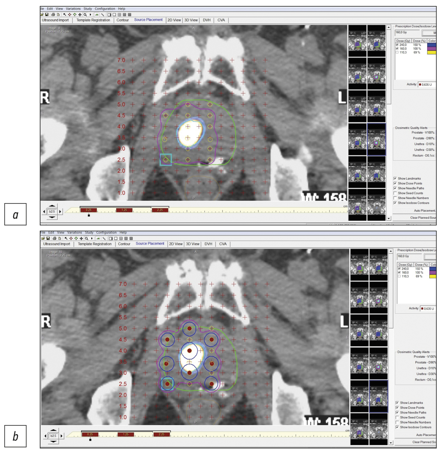
В данной работе на 7 клинических наблюдениях локализованного рака предстательной железы (шкала Глисона 6–7) показано, что прецизионность низкодозной брахитерапии микроисточниками 125I локализованных карцином предстательной железы, равно как и прицельной биопсии, может быть повышена при использовании гибридных методов ПСМА-рецепторной (простатспецифический мембранный антиген) молекулярной визуализации (ОФЭКТ/КТ, ПЭТ/КТ). Метод ОФЭКТ/КТ более доступен, чем ПЭТ/КТ, и при наличии холодных наборов (HYNIC-PSMA) позволяет выполнять исследование в любой лаборатории радиоизотопной диагностики, располагающей соответствующими сканерами.
Инновационная технология ПСМА-навигационной биопсии и брахитерапии под контролем гибридной молекулярной визуализации может применяться при первичных и рецидивных случаях локализованного рака предстательной железы, увеличивает точность и снижает травматичность процедур, повышает медико-экономическую эффективность низкодозной брахитерапии микроисточниками 125I.
Необходимы дальнейшие исследования для совершенствования технологии и оценки отдалённых результатов лечения на многочисленной группе пациентов.
Полный текст
Открыть статью на сайте журналаОб авторах
Павел Владимирович Свиридов
Медицинский центр «Доктор Плюс»
Email: p_sviridov73@mail.ru
ORCID iD: 0009-0008-3362-8255
SPIN-код: 4702-3067
Россия, Обнинск
Павел Олегович Румянцев
Группа клиник «Мой медицинский центр»
Автор, ответственный за переписку.
Email: pavelrum@gmail.com
ORCID iD: 0000-0002-7721-634X
SPIN-код: 7085-7976
Scopus Author ID: 110759
д-р мед. наук
Россия, Санкт-ПетербургМихаил Владимирович Дегтярев
Национальный медицинский исследовательский центр эндокринологии
Email: germed@mail.ru
ORCID iD: 0000-0001-5652-2607
SPIN-код: 7725-7831
Россия, Москва
Сергей Сергеевич Серженко
Национальный медицинский исследовательский центр эндокринологии
Email: vv1ld@yandex.ru
ORCID iD: 0000-0003-2326-1396
SPIN-код: 4713-8986
Россия, Москва
Дмитрий Борисович Санин
Медицинский центр «Доктор Плюс»; Национальный медицинский исследовательский центр радиологии
Email: dimitresko82@yandex.ru
ORCID iD: 0009-0004-2047-4921
SPIN-код: 8939-9101
канд. биол. наук
Россия, Обнинск; ОбнинскСергей Викторович Стыров
Медицинский центр «Доктор Плюс»
Email: rizost@yandex.ru
ORCID iD: 0000-0003-4315-8855
SPIN-код: 9019-8520
Scopus Author ID: 924845
Россия, Обнинск
Дмитрий Юрьевич Агибалов
Медицинский центр «Доктор Плюс»
Email: agibalovd@bk.ru
ORCID iD: 0000-0003-2995-7140
SPIN-код: 6938-5804
Россия, Обнинск
Сергей Владимирович Коренев
Балтийский федеральный университет имени Иммануила Канта
Email: korenevsv@mail.ru
ORCID iD: 0000-0003-2310-0576
SPIN-код: 5257-4476
д-р мед. наук, профессор
Россия, КалининградСписок литературы
- Sung H., Ferlay J., Siegel R.L., et al. Global Cancer Statistics 2020: GLOBOCAN Estimates of Incidence and Mortality Worldwide for 36 Cancers in 185 Countries // CA Cancer J Clin. 2021. Vol. 71, N 3. Р. 209–249. doi: 10.3322/caac.21660
- Nyame Y.A., Gulati R., Tsodikov A., et al. Prostate-Specific antigen screening and recent increases in advanced prostate cancer // JNCI Cancer Spectr. 2021. Vol. 5, N 1. Р. pkaa. 098 doi: 10.1093/jncics/pkaa098
- Pommier P., Ferré M., Blanchard P., et al. Prostate cancer brachytherapy: SFRO guidelines 2021 // Cancer Radiotherap. 2022. Vol. 26, N 1-2. Р. 344–355. doi: 10.1016/j.canrad.2021.11.019
- Parker C., Castro E., Fizazi K., et al. Prostate cancer: ESMO Clinical Practice Guidelines for diagnosis, treatment and follow-up // Ann Oncol. 2020. Vol. 31, N 9. Р. 1119–1134. doi: 10.1016/j.annonc.2020.06.011
- Mottet N., van der Berg R., Briers E., et al. EAU-eanm-estro-esur-siog guidelines on prostate cancer-2020 update. Part 1: Screening, diagnosis, and local treatment with curative intent // Eur Urol. 2021. Vol. 79, N 2. Р. 243–262. doi: 10.1016/j.eururo.2020.09.042
- Носов Д.А., Волкова М.И., Гладков О.А., и др. Практические рекомендации по лечению рака предстательной железы // Злокачественные опухоли. Практические рекомендации RUSSCO. 2022. Т. 12, № #3s2. С. 607–626. doi: 10.18027/2224-5057-2022-12-3s2-607-626
- Tsumura H., Tanaka N., Oguchi T., et al. Comparative effectiveness of low-dose-rate brachytherapy with or without external beam radiotherapy in favorable and unfavorable intermediate-risk prostate cancer // Sci Rep. 2022. Vol. 12, N 1. Р. 11023. doi: 10.1038/s41598-022-15028-6
- Tanaka N., Asakawa I., Hasegawa M., Fujimoto K. Low-dose-rate brachytherapy for prostate cancer: A 15-year experience in Japan // Int J Urol. 2020. Vol. 27, N 1. Р. 17–23. doi: 10.1111/iju.14098
- Fellin G., Mirri M.A., Santoro L., et al. Low dose rate brachytherapy (LDR-BT) as monotherapy for early stage prostate cancer in Italy: Practice and outcome analysis in a series of 2237 patients from 11 institutions // Br J Radiol. 2016. Vol. 89, N 1065. Р. 20150981. doi: 10.1259/bjr.20150981
- Okamoto K., Okuyama K., Kohno N., Tsugawa T. Clinical outcomes of low-dose-rate brachytherapy based radiotherapy for intermediate risk prostate cancer // J Contemp Brachytherapy. 2020. Vol. 12, N 1. Р. 6–11. doi: 10.5114/jcb.2020.92405
- Cunha J.A., Flynn R., Bélanger C., et al. Brachytherapy future directions // Semin Radiat Oncol. 2020. Vol. 30, N 1. Р. 94–106. doi: 10.1016/j.semradonc.2019.09.001
- Afshar-Oromieh A. PSMA-ligand imaging in the diagnosis of prostate cancer // Clinical Nuclear Medicine: Second Edition. Springer International Publishing, 2020. Р. 755–763. doi: 10.1007/978-3-030-39457-8_25
- Zippel C., Ronski S.C., Bohnet-Joschko S., et al. Current status of PSMA-radiotracers for prostate cancer: Data analysis of prospective trials listed on clinicaltrials.gov // Pharmaceuticals. 2020. Vol. 13, N 1. Р. 12. doi: 10.3390/ph13010012
- Зырянов А.В., Ощепков В.Н., Свиридов П.В., и др. Рекомендации по лечению рака предстательной железы с помощью низкодозной перманентной внутритканевой лучевой терапии (брахитерапии). Экспертное совещание Объединения брахитерапевтов России (ОБР), 4 октября 2014, Москва // Экспериментальная и клиническая урология. 2015. № 2. С. 37–46.
- Kasivisvanathan V., Rannikko A.S., Borghi M., et al. MRI-Targeted or standard biopsy for prostate-cancer diagnosis // N Engl J Med. 2018. Vol. 378, N 19. Р. 1767–1777. doi: 10.1056/nejmoa1801993
- Sazuka T., Imamoto T., Namekawa T., et al. Analysis of preoperative detection for apex prostate cancer by transrectal biopsy // Prostate Cancer. 2013. Vol. 2013. Р. 705865. doi: 10.1155/2013/705865
- Tewes S., Peters I., Tiemeyer A., et al. Evaluation of MRI/ Ultrasound fusion-guided prostate biopsy using transrectal and transperineal approaches // Biomed Res Int. 2017. Vol. 2017. Р. 2176471. doi: 10.1155/2017/2176471
- Qiu D.X., Li J., Zhang J.W., et al. Dual-tracer PET/CT-targeted, mpMRI-targeted, systematic biopsy, and combined biopsy for the diagnosis of prostate cancer: A pilot study // Eur J Nucl Med Mol Imaging. 2022. Vol. 49, N 8. Р. 2821–2832. doi: 10.1007/s00259-021-05636-1
- Donato P., Morton A., Yaxley J., et al. 68Ga-PSMA PET/CT better characterizes localised prostate cancer after MRI and transperineal prostate biopsy: Is 68Ga-PSMA PET/CT guided biopsy the future? // Eur J Nucl Med Mol Imaging. 2020. Vol. 47, N 8. Р. 1843–1851. doi: 10.1007/s00259-019-04620-0
- Zhang L.L., Li W.C., Xu Z., et al. 68Ga-PSMA PET/CT targeted biopsy for the diagnosis of clinically significant prostate cancer compared with transrectal ultrasound guided biopsy: A prospective randomized single-centre study // Eur J Nucl Med Mol Imaging. 2021. Vol. 48, N 2. Р. 483–492 doi: 10.1007/s00259-020-04863-2
- Duan H., Ghanouni P., Daniel B., et al. A pilot study of 68Ga-PSMA11 and 68Ga-RM2 PET/MRI for biopsy guidance in patients with suspected prostate cancer // J Med. 2022. Vol. 64, N 5. Р. 744–750. doi: 10.2967/jnumed.122.264448
- Chin J., Rumble R.B., Kollmeier M., et al. Brachytherapy for patients with prostate cancer: American Society of Clinical Oncology / Cancer Care Ontario joint guideline update // J Clin Oncol. 2017. Vol. 35, N 15. Р. 1737–1745. doi: 10.1200/JCO.2016.72.0466
- Basu S., Alavi A. SPECT-CT and PET-CT in oncology: An overview // Curr Med Imaging Rev. 2011. Vol. 7, N 3. Р. 202–209. doi: 10.2174/157340511796411168
- Soldatov A., von Klot C.A., Walacides D., et al. Patterns of progression after 68Ga-PSMA-ligand PET/CT-guided radiation therapy for recurrent prostate cancer // Int J Radiat Oncol Biol Phys. 2019. Vol. 103, N 1. Р. 95–104. doi: 10.1016/j.ijrobp.2018.08.066
- Werner P., Neumann C., Eiber M., et al. [99cmTc]Tc-PSMA-I&S-SPECT/CT: experience in prostate cancer imaging in an outpatient center // EJNMMI Res. 2020. Vol. 10, N 1. Р. 45. doi: 10.1186/s13550-020-00635-z
- Berliner C., Steinhelfer L., Chantadisai M., et al. Delayed imaging improves lesion detectability in [99mTc]Tc-PSMA-I&S SPECT/CT in recurrentprostate cancer // J Nucl Med. 2023. Vol. 64, N 7. Р. 1036–1042. doi: 10.2967/jnumed.122.265252
- Румянцев П.О. Возрастающая роль методов функциональной визуализации для навигации дистанционной радиотерапии и брахитерапии на примере рака предстательной железы // Digital Diagnostics. 2022. Т. 2, № 4. С. 488–497. doi: 10.17816/DD96197
- Патент РФ на изобретение № RU 2788859 С2. Агибалов Д.Ю., Дегтярев М.В., Румянцев П.О., и др. Способ прицельной брахитерапии рака предстательной железы под навигацией гибридной ПСМА-рецепторной сцинтиграфии. Режим доступа: https://yandex.ru/patents/doc/RU2788859C2_20230125. Дата обращения: 15.08.2023.
Дополнительные файлы


