

")


在PSMA受体分子成像控制下的前列腺癌精确近距离照射
- 作者: Sviridov P.V.1, Rumiantsev P.O.2, Degtyarev M.V.3, Serzhenko S.S.3, Sanin D.B.1,4, Styrov S.V.1, Agibalov D.Y.1, Korenev S.V.5
-
隶属关系:
- Medical center “Doctor Plus”
- Clinics group “My Medical Center”
- Endocrinology Research Centre
- National Medical Research Radiological Center
- I. Kant Baltic Federal University
- 期: 卷 4, 编号 3 (2023)
- 页面: 411-426
- 栏目: 临床病例及临床病例的系列
- URL: https://ogarev-online.ru/DD/article/view/254079
- DOI: https://doi.org/10.17816/DD340815
- ID: 254079
如何引用文章

详细
治疗无腺囊萌发迹象和无转移迹象(cT1-T2N0M0期)的局部前列腺癌的方法之一是植入基于同位素125I的微源近距离照射。结构成像方法(超声检查、电子计算机断层扫描(CT)、磁共振成像(MRI))在前列腺癌的鉴别诊断中特异性不高。混合放射成像技术(单光子发射计算机断层扫描+电子计算机断层扫描,SPECT/CT;正电子发射计算机断层扫描+电子计算机断层扫描,PET/CT;正电子发射断层扫描+磁共振成像,PET/MRI)结合了结构成像的横断面方法(CT和MRI)的高灵敏度和分子成像方法(SPECT、PET)的高特异性与肿瘤放射治疗物的优点。
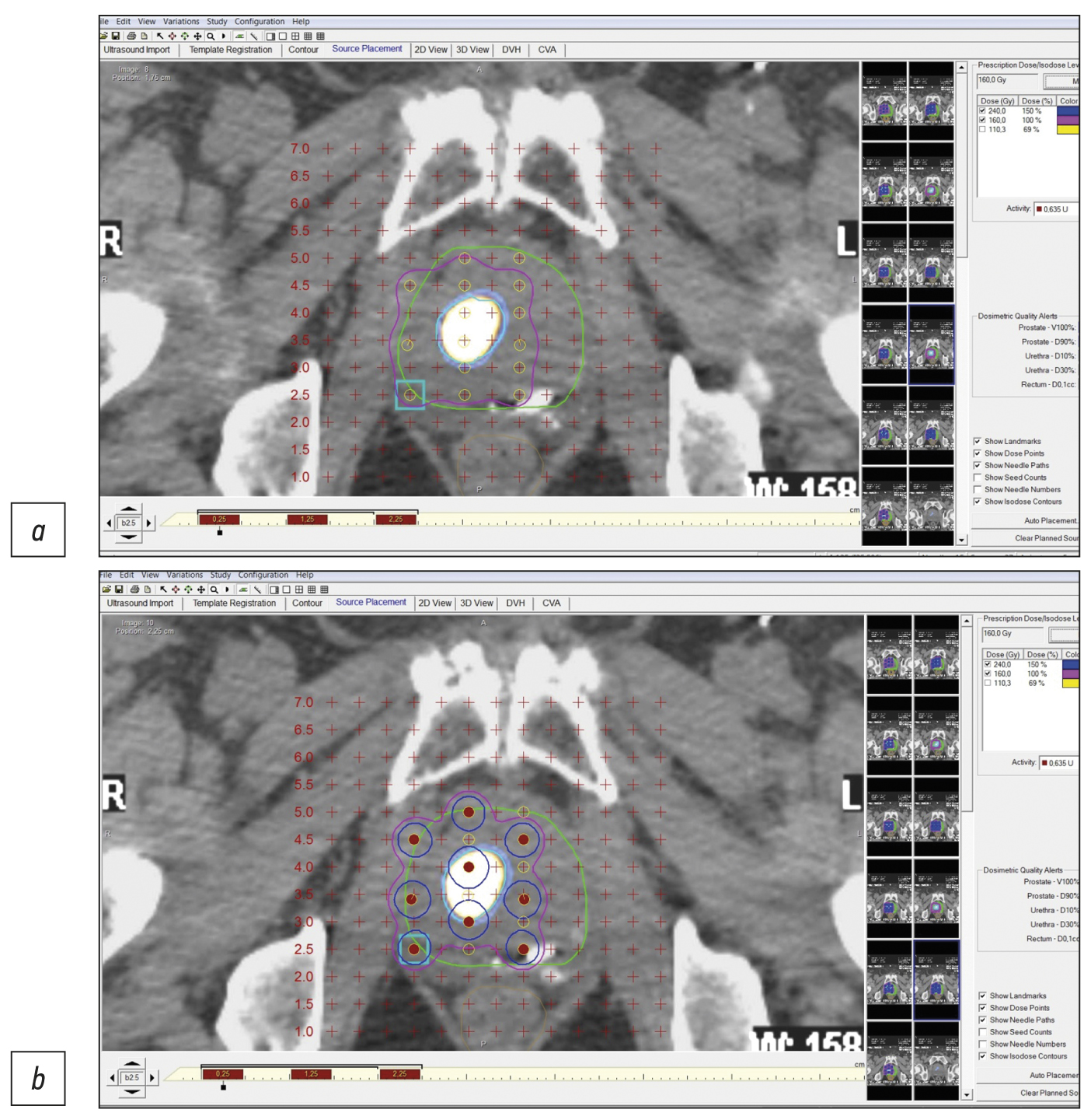
在这项研究中,7例局部前列腺癌(格里森评分为6-7分)的临床观察结果表明了,通过使用PSMA受体(前列腺特异性膜抗原)分子成像(SPECT/CT、PET/CT)的混合方法,可以提高使用微源125I对局部前列腺癌进行低剂量近距离照射以及靶向活检的精确度。SPECT/CT比PET/CT更经济实惠,而且在存在冷试剂盒(HYNIC-PSMA)的情况下,允许在任何拥有适当扫描仪的放射性同位素诊断实验室进行检查。
混合分子成像控制下的PSMA引导活检和近距离照射创新技术可用于原发性和复发性的局部前列腺癌病例,提高准确度,减少检查创伤性,提高微源125I低剂量近距离照射的医疗和经济效益。
还需要进一步的研究来改进这种技术,并对大量患者的长期治疗效果进行评估。
作者简介
Pavel V. Sviridov
Medical center “Doctor Plus”
Email: p_sviridov73@mail.ru
ORCID iD: 0009-0008-3362-8255
SPIN 代码: 4702-3067
俄罗斯联邦, Obninsk
Pavel O. Rumiantsev
Clinics group “My Medical Center”
编辑信件的主要联系方式.
Email: pavelrum@gmail.com
ORCID iD: 0000-0002-7721-634X
SPIN 代码: 7085-7976
Scopus 作者 ID: 110759
MD, Dr. Sci. (Med.)
俄罗斯联邦, Saint PeterburgMikhail V. Degtyarev
Endocrinology Research Centre
Email: germed@mail.ru
ORCID iD: 0000-0001-5652-2607
SPIN 代码: 7725-7831
俄罗斯联邦, Moscow
Sergey S. Serzhenko
Endocrinology Research Centre
Email: vv1ld@yandex.ru
ORCID iD: 0000-0003-2326-1396
SPIN 代码: 4713-8986
俄罗斯联邦, Moscow
Dmitry B. Sanin
Medical center “Doctor Plus”; National Medical Research Radiological Center
Email: dimitresko82@yandex.ru
ORCID iD: 0009-0004-2047-4921
SPIN 代码: 8939-9101
Cand. Sci. (Biol.)
俄罗斯联邦, Obninsk; ObninskSergey V. Styrov
Medical center “Doctor Plus”
Email: rizost@yandex.ru
ORCID iD: 0000-0003-4315-8855
SPIN 代码: 9019-8520
Scopus 作者 ID: 924845
俄罗斯联邦, Obninsk
Dmitry Yu. Agibalov
Medical center “Doctor Plus”
Email: agibalovd@bk.ru
ORCID iD: 0000-0003-2995-7140
SPIN 代码: 6938-5804
俄罗斯联邦, Obninsk
Sergey V. Korenev
I. Kant Baltic Federal University
Email: korenevsv@mail.ru
ORCID iD: 0000-0003-2310-0576
SPIN 代码: 5257-4476
MD, Dr. Sci. (Med.), Professor
俄罗斯联邦, Kaliningrad参考
- Sung H, Ferlay J, Siegel RL, et al. Global cancer statistics 2020: Globocan estimates of incidence and mortality worldwide for 36 cancers in 185 countries. CA Cancer J Clin. 2021;71(3):209–249. doi: 10.3322/caac.21660
- Nyame YA, Gulati R, Tsodikov A, et al. Prostate-Specific antigen screening and recent increases in advanced prostate cancer. JNCI Cancer Spectr. 2021;5(1):pkaa098. doi: 10.1093/jncics/pkaa098
- Pommier P, Ferré M, Blanchard P, et al. Prostate cancer brachytherapy: SFRO guidelines 2021. Cancer Radiotherap. 2022;26(1-2):344–355. doi: 10.1016/j.canrad.2021.11.019
- Parker C, Castro E, Fizazi K, et al. Prostate cancer: ESMO clinical practice guidelines for diagnosis, treatment and follow-up. Ann Oncol. 2020;31(9):1119–1134. doi: 10.1016/j.annonc.2020.06.011
- Mottet N, van der Berg R, Briers E, et al. EAU-eanm-estro-esur-siog guidelines on prostate cancer-2020 update. Part 1: Screening, diagnosis, and local treatment with curative intent. Eur Urol. 2021;79(2):243–262. doi: 10.1016/j.eururo.2020.09.042
- Nosov DA, Volkova MI, Gladkov OA, et al. Practical recommendations for the treatment of prostate cancer. Malignant Tumors. Practical recommendations RUSSCO. 2022;12(#3s2):607–626. (In Russ). doi: 10.18027/2224-5057-2022-12-3s2-607-626
- Tsumura H, Tanaka N, Oguchi T, et al. Comparative effectiveness of low-dose-rate brachytherapy with or without external beam radiotherapy in favorable and unfavorable intermediate-risk prostate cancer. Sci Rep. 2022;12(1):11023. doi: 10.1038/s41598-022-15028-6
- Tanaka N, Asakawa I, Hasegawa M, Fujimoto K. Low-dose-rate brachytherapy for prostate cancer: A 15-year experience in Japan. Int J Urol. 2020;27(1):17–23. doi: 10.1111/iju.14098
- Fellin G, Mirri MA, Santoro L, et al. Low dose rate brachytherapy (LDR-BT) as monotherapy for early stage prostate cancer in Italy: Practice and outcome analysis in a series of 2237 patients from 11 institutions. Br J Radiol. 2016;89(1065):20150981. doi: 10.1259/bjr.20150981
- Okamoto K, Okuyama K, Kohno N, Tsugawa T. Clinical outcomes of low-dose-rate brachytherapy based radiotherapy for intermediate risk prostate cancer. J Contemp Brachytherapy. 2020;12(1):6–11. doi: 10.5114/jcb.2020.92405
- Cunha JA, Flynn R, Bélanger C, et al. Brachytherapy future directions. Semin Radiat Oncol. 2020;30(1):94–106. doi: 10.1016/j.semradonc.2019.09.001
- Afshar-Oromieh A. PSMA-ligand imaging in the diagnosis of prostate cancer. In: Clinical Nuclear Medicine: Second Edition. Springer International Publishing; 2020. Р. 755–763. doi: 10.1007/978-3-030-39457-8_25
- Zippel C, Ronski SC, Bohnet-Joschko S, et al. Current status of PSMA-radiotracers for prostate cancer: Data analysis of prospective trials listed on clinicaltrials.gov. Pharmaceuticals. 2020;13(1):12. doi: 10.3390/ph13010012
- Zyryanov AV, Oshchepkov VN, Sviridov PV, et al. Recommendations for the treatment of prostate cancer with low-dose permanent interstitial radiation therapy (brachytherapy). Expert meeting of the Association of Brachytherapists of Russia (OBR), October 4, 2014, Moscow. Experimental Clin Urol. 2015;(2):37–46. (In Russ).
- Kasivisvanathan V, Rannikko AS, Borghi M, et al. MRI-targeted or standard biopsy for prostate-cancer diagnosis. N Engl J Med. 2018;378(19):1767–1777 doi: 10.1056/nejmoa1801993
- Sazuka T, Imamoto T, Namekawa T, et al. Analysis of preoperative detection for apex prostate cancer by transrectal biopsy. Prostate Cancer. 2013;2013:705865. doi: 10.1155/2013/705865
- Tewes S, Peters I, Tiemeyer A, et al. Evaluation of MRI/ Ultrasound fusion-guided prostate biopsy using transrectal and transperineal approaches. Biomed Res Int. 2017;2017:2176471. doi: 10.1155/2017/2176471
- Qiu DX, Li J, Zhang JW, et al. Dual-tracer PET/CT-targeted, mpMRI-targeted, systematic biopsy, and combined biopsy for the diagnosis of prostate cancer: A pilot study. Eur J Nucl Med Mol Imaging. 2022;49(8):2821–2832. doi: 10.1007/s00259-021-05636-1
- Donato P, Morton A, Yaxley J, et al. 68Ga-PSMA PET/CT better characterizes localised prostate cancer after MRI and transperineal prostate biopsy: Is 68Ga-PSMA PET/CT guided biopsy the future? Eur J Nucl Med Mol Imaging. 2020;47(8):1843–1851. doi: 10.1007/s00259-019-04620-0
- Zhang LL, Li WC, Xu Z, et al. 68Ga-PSMA PET/CT targeted biopsy for the diagnosis of clinically significant prostate cancer compared with transrectal ultrasound guided biopsy: A prospective randomized single-centre study. Eur J Nucl Med Mol Imaging. 2021;48(2):483–492. doi: 10.1007/s00259-020-04863-2
- Duan H, Ghanouni P, Daniel B, et al. A pilot study of 68Ga-PSMA11 and 68Ga-RM2 PET/MRI for biopsy guidance in patients with suspected prostate cancer. J Nuclear Med. 2022;64(5):744–750. doi: 10.2967/jnumed.122.264448
- Chin J, Rumble RB, Kollmeier M, et al. Brachytherapy for patients with prostate cancer: American Society of Clinical Oncology / Cancer Care Ontario joint guideline update. J Clin Oncol. 2017;35(15):1737–1745. doi: 10.1200/JCO.2016.72.0466
- Basu S, Alavi A. SPECT-CT and PET-CT in oncology: An overview. Curr Med Imaging Rev. 2011;7(3):202–209. doi: 10.2174/157340511796411168
- Soldatov A, von Klot CA, Walacides D, et al. Patterns of progression after 68Ga-PSMA-Ligand PET/CT-Guided radiation therapy for recurrent prostate cancer. Int J Radiat Oncol Biol Phys. 2019;103(1):95–104. doi: 10.1016/j.ijrobp.2018.08.066
- Werner P, Neumann C, Eiber M, et al. [99cmTc]Tc-PSMA-I&S-SPECT/CT: experience in prostate cancer imaging in an outpatient center. EJNMMI Res. 2020;10(1):45. doi: 10.1186/s13550-020-00635-z
- Berliner C, Steinhelfer L, Chantadisai M, et al. Delayed imaging improves lesion detectability in [99mTc]Tc-PSMA-I&S SPECT/CT in recurrentprostate cancer. J Nucl Med. 2023;64(7):1036–1042. doi: 10.2967/jnumed.122.265252
- Rumyantsev PO. The increasing role of functional imaging methods for navigation of remote radiotherapy and brachytherapy on the example of prostate cancer. Digital Diagnostics. 2022;2(4):488–497. (In Russ). doi: 10.17816/DD96197
- Patent RUS № RU 2788859 С2. Agibalov DYu, Degtyarev MV, Rumyantsev PO, et al. Method of targeted brachytherapy of prostate cancer under the navigation of hybrid PSMA-receptor scintigraphy. Available from: https://yandex.ru/patents/doc/RU2788859C2_20230125. Accessed: 15.08.2023.
补充文件


