

")


Machine-learning and artificial neural network technologies in the classification of postkeratotomic corneal deformity
- Авторлар: Tsyrenzhapova E.K.1, Rozanova O.I.1, Iureva T.N.1,2,3, Ivanov A.A.1, Rozanov I.S.4
-
Мекемелер:
- The S. Fyodorov Eye Microsurgery Federal State Institution
- Irkutsk State Medical University
- Russian Medical Academy of Continuous Professional Education
- LLC Transneft Technology
- Шығарылым: Том 5, № 1 (2024)
- Беттер: 64-74
- Бөлім: Original Study Articles
- URL: https://ogarev-online.ru/DD/article/view/262961
- DOI: https://doi.org/10.17816/DD624022
- ID: 262961
Дәйексөз келтіру

Аннотация
BACKGROUND: A thorough analysis of both optical and anatomical properties of the cornea in patients after anterior radial keratotomy is important in choosing the optical power of an intraocular lens in the surgical treatment of cataracts and other types of optical correction. Improving the classification of postkeratotomic corneal deformity is crucial in modern ophthalmology due to its diverse clinical presentation.
AIM: To develop an automated classification system for postkeratotomic corneal deformity using machine learning and artificial neural networks based on the analysis of topographic maps of the cornea.
MATERIALS AND METHODS: Depersonalized data from medical records of 250 patients aged 46–76 (mean, 59.63±5.95) years were analyzed. Moreover, 500 topographic maps of the anterior and posterior surfaces of the cornea were analyzed, and three stages of machine learning for postkeratotomic corneal deformity classification were performed.
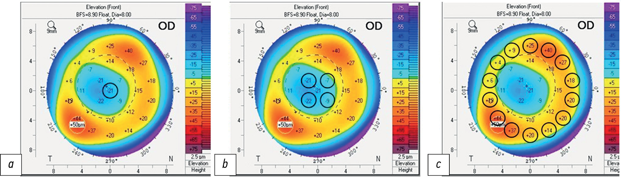
RESULTS: Stage I, which involved topography analysis of the anterior and posterior surfaces of the cornea, allowed for the measurement of anterior and posterior corneal elevation in three ring-shaped zones. At stage II, a direct distribution neural network was selected and created during deep machine learning. Eight auxiliary parameters describing the shape of the anterior and posterior surfaces of the cornea were established. In Stage III, classification algorithms for postkeratotomic corneal deformity were developed based on the test-to-training sample ratio, which ranged from 75% to 91%.
CONCLUSION: The proposed artificial neural network classifies postkeratotomic corneal deformity types with an accuracy of 91%. The potential for further improving the training quality of this artificial neural network has been established. Neural network algorithms can become a useful tool for the automatic classification of postkeratotomic corneal deformity in patients after radial keratotomy.
Негізгі сөздер
Толық мәтін
##article.viewOnOriginalSite##Авторлар туралы
Ekaterina Tsyrenzhapova
The S. Fyodorov Eye Microsurgery Federal State Institution
Email: katyakel@mail.ru
ORCID iD: 0000-0002-6804-8268
SPIN-код: 1158-5233
MD
Ресей, IrkutskOlga Rozanova
The S. Fyodorov Eye Microsurgery Federal State Institution
Email: olgrozanova@gmail.com
ORCID iD: 0000-0003-3139-2409
SPIN-код: 6557-9123
MD, Dr. Sci. (Medicine)
Ресей, IrkutskTatiana Iureva
The S. Fyodorov Eye Microsurgery Federal State Institution; Irkutsk State Medical University; Russian Medical Academy of Continuous Professional Education
Email: tnyurieva@mail.ru
ORCID iD: 0000-0003-0547-7521
SPIN-код: 8457-5851
MD, Dr. Sci. (Medicine), Professor
Ресей, Irkutsk; Irkutsk; IrkutskAndrey Ivanov
The S. Fyodorov Eye Microsurgery Federal State Institution
Email: ivanov.andrei.med@yandex.ru
ORCID iD: 0009-0001-4235-9252
MD
Ресей, IrkutskIvan Rozanov
LLC Transneft Technology
Хат алмасуға жауапты Автор.
Email: nauka@mntk.irkutsk.ru
ORCID iD: 0009-0001-7202-0428
Ресей, Irkutsk
Әдебиет тізімі
- Issarti I, Consejo A, Jiménez-García M, et al. Computer aided diagnosis for suspect keratoconus detection. Comput Biol Med. 2019;109:33–42. doi: 10.1016/j.compbiomed.2019.04.024
- Chen X, Zhao J, Iselin KC, et al. Keratoconus detection of changes using deep learning of colour-coded maps. BMJ Open Ophthalmol. 2021;6(1):e000824. doi: 10.1136/bmjophth-2021-000824
- Feng R, Xu Z, Zheng X, et al. KerNet: A novel deep learning approach for keratoconus and sub-clinical keratoconus detection based on raw data of the pentacam HR system. IEEE J Biomed Health Inform. 2021;25(10):3898–3910. doi: 10.1109/JBHI.2021.3079430
- Gatinel D. Screening for subclinical keratoconus and prevention of corneal ectasia with SCORE analyzer software. In: Febbraro J-L, Khan HN, Koch DD, editors. Surgical correction of astigmatism. Cham: Springer International Publishing; 2018. doi: 10.1007/978-3-319-56565-1_9
- Ruiz Hidalgo I, Rozema JJ, Saad A, et al. Validation of an objective keratoconus detection system implemented in a scheimpflug tomographer and comparison with other methods. Cornea. 2017;36(6):689–695. doi: 10.1097/ICO.0000000000001194
- Malyugin BE, Sakhnov SN, Axenova LE, Myasnikova VV. Application of artificial intelligence in diagnostics and surgery of keratoconus: a systematic overview. Fyodorov Journal of Ophthalmic Surgery. 2022;(1):77–96. EDN: PPQRWZ doi: 10.25276/0235-4160-2022-1-77-96
- Abdelmotaal H, Mostafa MM, Mostafa ANR, et al. Classification of Color-Coded Scheimpflug Camera Corneal Tomography Images Using Deep Learning. Transl Vis Sci Technol. 2020;9(13):30. doi: 10.1167/tvst.9.13.30
- Dos Santos VA, Schmetterer L, Stegmann H, et al. CorneaNet: fast segmentation of cornea OCT scans of healthy and keratoconic eyes using deep learning. Biomed Opt Express. 2019;10(2):622–641. doi: 10.1364/BOE.10.000622
- Kuo BI, Chang WY, Liao TS, et al. Keratoconus Screening Based on Deep Learning Approach of Corneal Topography. Transl Vis Sci Technol. 2020;9(2):53. doi: 10.1167/tvst.9.2.53
- Shi C, Wang M, Zhu T, et al. Machine learning helps improve diagnostic ability of subclinical keratoconus using Scheimpflug and OCT imaging modalities. Eye Vis (Lond). 2020;7:48. doi: 10.1186/s40662-020-00213-3
- Shukhaev SV, Mordovtseva EA, Pustozerov EA, Kudlakhmedov SS Application of convolutional neural networks to define Fuchs endothelial dystrophy. Fyodorov Journal of Ophthalmic Surgery. 2022;(S4):70–76. EDN: WEZTKV doi: 10.25276/0235-4160-2022-4S-70-76
- Obaid HS, Dheyab SA, Sabry SS. The impact of data pre-processing techniques and dimensionality reduction on the accuracy of machine learning. 2019 9th Annu. Inf. Technol. Electromechanical Eng. Microelectron. Conf. IEMECON. 2019:279–283. doi: 10.1109/IEMECONX.2019.8877011
- Valdés-Mas MA, Martín-Guerrero JD, Rupérez MJ, et al. A new approach based on Machine Learning for predicting corneal curvature (K1) and astigmatism in patients with keratoconus after intracorneal ring implantation. Comput Methods Programs Biomed. 2014;116:39–47. doi: 10.1016/j.cmpb.2014.04.003
- Patent RUS № RU 2793142 C1/ 29.03.2023. Rozanova OI, Tsyrenzhapova EK, Iureva TN, et al. A method of evaluating the relief of the anterior and posterior corneal surface. (In Russ).
- Arbelaez MC, Versaci F, Vestri G, et al. Use of a Support Vector Machine for Keratoconus and Subclinical Keratoconus Detection by Topographic and Tomographic Data. Ophthalmology. 2012;119(11):2231–2238. doi: 10.1016/j.ophtha.2012.06.005
- Ruiz Hidalgo I, Rodriguez P, Rozema JJ, et al. Evaluation of a Machine-Learning Classifier for Keratoconus Detection Based on Scheimpflug Tomography. Cornea. 2016;35(6):827–832. doi: 10.1097/ico.0000000000000834
Қосымша файлдар


