

")


Диагностика внутричерепных кровоизлияний по данным компьютерной томографии головного мозга с помощью искусственного интеллекта
- Авторы: Хоружая А.Н.1, Арзамасов К.М.1, Коденко М.Р.1, Кремнёва Е.И.1,2, Буренчев Д.В.1
-
Учреждения:
- Научно-практический клинический центр диагностики и телемедицинских технологий
- Российский центр неврологии и нейронаук
- Выпуск: Том 6, № 2 (2025)
- Страницы: 214-228
- Раздел: Оригинальные исследования
- URL: https://ogarev-online.ru/DD/article/view/310211
- DOI: https://doi.org/10.17816/DD645364
- EDN: https://elibrary.ru/RFYVMC
- ID: 310211
Цитировать
Полный текст

Аннотация
Обоснование. Внутричерепные кровоизлияния характеризуются высокой летальностью и риском инвалидизации, что обусловливает необходимость оперативной и точной диагностики, особенно в первые 24 часа. Использование технологий искусственного интеллекта для анализа компьютерных томограмм головного мозга позволяет сократить время диагностики и улучшить её качество. Актуальность работы подчёркнута ограниченным числом сертифицированных в России сервисов искусственного интеллекта для выявления внутричерепных кровоизлияний, а также отсутствием данных о их долгосрочной эффективности, что обусловливает необходимость многоцентрового мониторинга для оценки устойчивости и точности таких систем в реальной клинической практике.
Цель исследования. Оценить диагностическую точность и устойчивость сервиса искусственного интеллекта для диагностики внутричерепных кровоизлияний по данным нативной компьютерной томографии головного мозга в условиях многоцентрового клинического мониторинга на протяжении 18 месяцев.
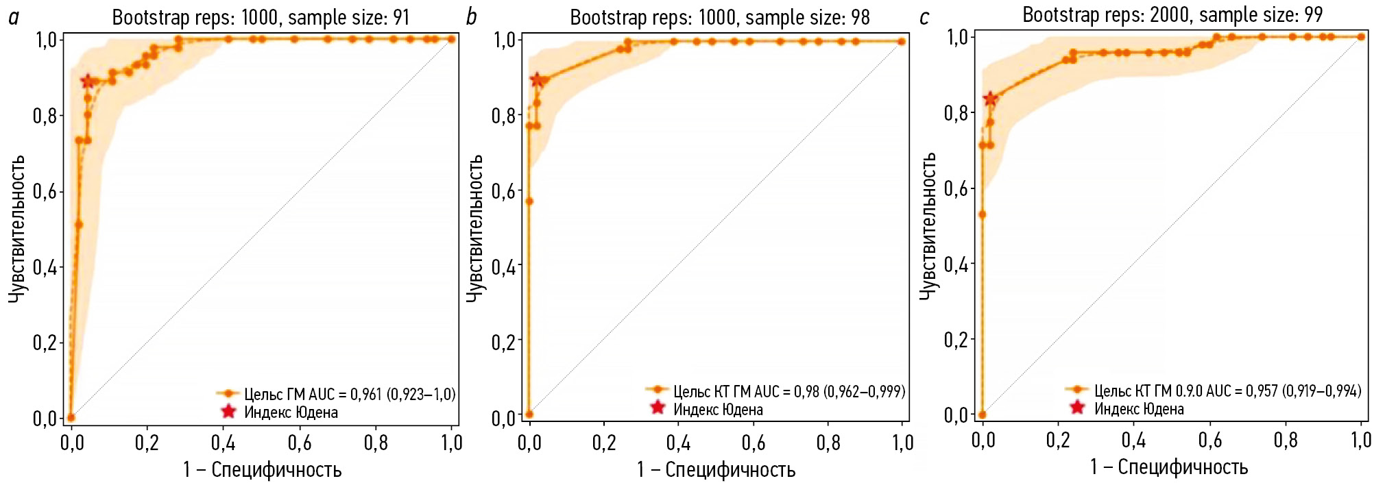
Методы. Для анализа использовали анонимизированные компьютерные томограммы головного мозга. Сервис искусственного интеллекта прошёл трёхэтапное тестирование для оценки его точности и клинической производительности на ограниченных наборах данных. В течение 18 месяцев два врача-рентгенолога, специализирующиеся на нейровизуализации, ежемесячно оценивали 80 компьютерно-томографических исследований головного мозга, предварительно обработанных сервисом искусственного интеллекта и случайным образом выбранных из клинического потока. Результаты проанализировали методом ROC-анализа с вычислением таких метрик, как чувствительность, специфичность, точность, площадь под характеристической кривой.
Результаты. При клиническом мониторинге проанализировано 1200 компьютерных томограмм головного мозга, из которых признаки внутричерепного кровоизлияния выявлены в 48,3% случаев. По результатам их бинарной классификации на наличие внутричерепных кровоизлияний, выполненной сервисом искусственного интеллекта, получены следующие диагностические метрики: чувствительность — 97,4% (95,8–98,5), специфичность — 75,4% (71,8–78,7), точность — 86,0% (83,9–87,9), площадь под характеристической кривой — 94% (92,6–95,3). Со временем наблюдали статистически значимую умеренную положительную корреляция между большинством диагностических метрик и временным показателем, за исключением чувствительности, что обусловлено сменой версии сервиса. Однако полное совпадение разметки и описания с заключением врача в выявленных сервисом искусственного интеллекта случаях внутричерепного кровоизлияния достигнуто в 28,5%, а различные расхождения найдены в 71,5%. Уточнённые метрики для случаев с полным соответствием заключения врача составили: чувствительность, специфичность, точность и площадь под характеристической кривой — 26,6, 73,8, 50,1 и 49,6% соответственно.
Заключение. Текущая конфигурация сервиса искусственного интеллекта позволяет исключать кровоизлияние с очень высокой вероятностью, что может быть полезно для первичной сортировки пациентов в условиях неотложной помощи. Однако низкие значения уточнённых метрик указывают на значительные расхождения между заключениями рентгенологов и результатами сервиса в аспектах детальной интерпретации патологии.
Полный текст
Открыть статью на сайте журналаОб авторах
Анна Николаевна Хоружая
Научно-практический клинический центр диагностики и телемедицинских технологий
Автор, ответственный за переписку.
Email: KhoruzhayaAN@zdrav.mos.ru
ORCID iD: 0000-0003-4857-5404
SPIN-код: 7948-6427
MD
Россия, МоскваКирилл Михайлович Арзамасов
Научно-практический клинический центр диагностики и телемедицинских технологий
Email: ArzamasovK@zdrav.mos.ru
ORCID iD: 0000-0001-7786-0349
SPIN-код: 3160-8062
д-р мед. наук
Россия, МоскваМария Романовна Коденко
Научно-практический клинический центр диагностики и телемедицинских технологий
Email: KodenkoM@zdrav.mos.ru
ORCID iD: 0000-0002-0166-3768
SPIN-код: 5789-0319
канд. техн. наук
Россия, МоскваЕлена Игоревна Кремнёва
Научно-практический клинический центр диагностики и телемедицинских технологий; Российский центр неврологии и нейронаук
Email: KremnevaE@zdrav.mos.ru
ORCID iD: 0000-0001-9396-6063
SPIN-код: 8799-8092
д-р мед. наук
Россия, Москва; МоскваДмитрий Владимирович Буренчев
Научно-практический клинический центр диагностики и телемедицинских технологий
Email: BurenchevD@zdrav.mos.ru
ORCID iD: 0000-0003-2894-6255
SPIN-код: 2411-3959
д-р мед. наук
Россия, МоскваСписок литературы
- Li X, Zhang L, Wolfe CDA, Wang Y. Incidence and long-term survival of spontaneous intracerebral hemorrhage over time: a systematic review and meta-analysis. Frontiers in Neurology. 2022;13:819737. doi: 10.3389/fneur.2022.819737 EDN: MLOQRJ
- Hemorrhagic stroke: clinical guidelines. Moscow: Ministry of Health of the Russian Federation; 2022. (In Russ.) [cited 2024 Dec 12]. Available from: https://ruans.org/Text/Guidelines/hemorrhagic-stroke-2022.pdf
- Hostettler IC, Seiffge DJ, Werring DJ. Intracerebral hemorrhage: an update on diagnosis and treatment. Expert Review of Neurotherapeutics. 2019;19(7):679–694. doi: 10.1080/14737175.2019.1623671 EDN: JWSYUZ
- Woo D, Comeau ME, Venema SU, et al. Risk factors associated with mortality and neurologic disability after intracerebral hemorrhage in a racially and ethnically diverse cohort. JAMA Network Open. 2022;5(3):e221103. doi: 10.1001/jamanetworkopen.2022.1103 EDN: BVHNLU
- Yaghi S, Dibu J, Achi E, et al. Hematoma expansion in spontaneous intracerebral hemorrhage: predictors and outcome. International Journal of Neuroscience. 2014;124(12):890–893. doi: 10.3109/00207454.2014.887716
- Gong B, Khalvati F, Ertl-Wagner BB, Patlas MN. Artificial intelligence in emergency neuroradiology: current applications and perspectives. Diagnostic and Interventional Imaging. 2025;106(4):135–142. doi: 10.1016/j.diii.2024.11.002 EDN: DHXSGS
- Arbabshirani MR, Fornwalt BK, Mongelluzzo GJ, et al. Advanced machine learning in action: identification of intracranial hemorrhage on computed tomography scans of the head with clinical workflow integration. npj Digital Medicine. 2018;1(1):9. doi: 10.1038/s41746-017-0015-z EDN: BORIWC
- Seyam M, Weikert T, Sauter A, et al. Utilization of artificial intelligence–based intracranial hemorrhage detection on emergent noncontrast CT images in clinical workflow. Radiology: Artificial Intelligence. 2022;4(2):e210168. doi: 10.1148/ryai.210168 EDN: HEPSBX
- Davis MA, Rao B, Cedeno PA, et al. machine learning and improved quality metrics in acute intracranial hemorrhage by noncontrast computed tomography. Current Problems in Diagnostic Radiology. 2022;51(4):556–561. doi: 10.1067/j.cpradiol.2020.10.007 EDN: NHQFYC
- O’Neill TJ, Xi Y, Stehel E, et al. Active reprioritization of the reading worklist using artificial intelligence has a beneficial effect on the turnaround time for interpretation of head CT with intracranial hemorrhage. Radiology: Artificial Intelligence. 2021;3(2):e200024. doi: 10.1148/ryai.2020200024 EDN: LCDGTM
- Smorchkova AK, Khoruzhaya AN, Kremneva EI, Petryaikin AV. Machine learning technologies in CT-based diagnostics and classification of intracranial hemorrhages. Burdenko's Journal of Neurosurgery. 2023;87(2):85. doi: 10.17116/neiro20238702185EDN: JVZDST
- Yu KH, Kohane IS. Framing the challenges of artificial intelligence in medicine. BMJ Quality & Safety. 2018;28(3):238–241. doi: 10.1136/bmjqs-2018-008551
- Allen B, Dreyer K, Stibolt R, et al. Evaluation and real-world performance monitoring of artificial intelligence models in clinical practice: try it, buy it, check it. Journal of the American College of Radiology. 2021;18(11):1489–1496. doi: 10.1016/j.jacr.2021.08.022 EDN: NMKGVD
- Recht MP, Dewey M, Dreyer K, et al. Integrating artificial intelligence into the clinical practice of radiology: challenges and recommendations. European Radiology. 2020;30(6):3576–3584. doi: 10.1007/s00330-020-06672-5 EDN: WWDEXB
- Vasiliev YuA, Vlazimirskyy AV, Omelyanskaya OV, et al. Methodology for testing and monitoring artificial intelligence-based software for medical diagnostics. Digital Diagnostics. 2023;4(3):252–267. doi: 10.17816/DD321971 EDN: UEDORU
- Morozov SP, Vladzimirsky AV, Klyashtornyy VG, et al. Clinical acceptance of software based on artificial intelligence technologies (radiology). Moscow: Research and Practical Clinical Center for Diagnostics and Telemedicine Technologies; 2019. EDN: GWJIMI
- Morozov SP, Vladzimirsky AV, Andreychenko AE, et al. Regulations for the preparation of data sets with a description of approaches to the formation of a representative data sample. Moscow: Research and Practical Clinical Center for Diagnostics and Telemedicine Technologies; 2022. (In Russ.) EDN: XENAJE
- Chetverikov SF, Arzamasov KM, Andreichenko AE, et al. Approaches to sampling for quality control of artificial intelligence in biomedical research. Sovremennye tehnologii v medicine. 2023;15(2):19. doi: 10.17691/stm2023.15.2.02 EDN: FUKXYC
- Kodenko MR, Bobrovskaya TM, Reshetnikov RV, et al. Empirical approach to sample size estimation for testing of AI algorithms. Doklady Mathematics. 2024;110(S1):S62–S74. doi: 10.1134/S1064562424602063 EDN: VJHJRD
- Salehinejad H, Kitamura J, Ditkofsky N, et al. A real-world demonstration of machine learning generalizability in the detection of intracranial hemorrhage on head computerized tomography. Scientific Reports. 2021;11(1):17051. doi: 10.1038/s41598-021-95533-2 EDN: SXLMCH
- Zia A, Fletcher C, Bigwood S, et al. Retrospective analysis and prospective validation of an AI-based software for intracranial haemorrhage detection at a high-volume trauma centre. Scientific Reports. 2022;12(1):19885. doi: 10.1038/s41598-022-24504-y EDN: IWNBET
- Ginat DT. Analysis of head CT scans flagged by deep learning software for acute intracranial hemorrhage. Neuroradiology. 2019;62(3):335–340. doi: 10.1007/s00234-019-02330-w EDN: WTOITQ
- Voter AF, Meram E, Garrett JW, Yu JPJ. Diagnostic accuracy and failure mode analysis of a deep learning algorithm for the detection of intracranial hemorrhage. Journal of the American College of Radiology. 2021;18(8):1143–1152. doi: 10.1016/j.jacr.2021.03.005 EDN: GPJYDS
- McLouth J, Elstrott S, Chaibi Y, et al. Validation of a deep learning tool in the detection of intracranial hemorrhage and large vessel occlusion. Frontiers in Neurology. 2021;12:656112. doi: 10.3389/fneur.2021.656112 EDN: FFIXVV
- Kundisch A, Hönning A, Mutze S, et al. Deep learning algorithm in detecting intracranial hemorrhages on emergency computed tomographies. PLOS ONE. 2021;16(11):e0260560. doi: 10.1371/journal.pone.0260560 EDN: QPACKZ
- Del Gaizo AJ, Osborne TF, Shahoumian T, Sherrier R. Deep learning to detect intracranial hemorrhage in a national teleradiology program and the impact on interpretation time. Radiology: Artificial Intelligence. 2024;6(5):e240067. doi: 10.1148/ryai.240067 EDN: EHHAOO
- Pettet G, West J, Robert D, et al. A retrospective audit of an artificial intelligence software for the detection of intracranial haemorrhage used by a teleradiology company in the United Kingdom. BJR|Open. 2023;6(1):tzae033. doi: 10.1093/bjro/tzae033 EDN: DWNYCF
- Mäenpää SM, Korja M. Diagnostic test accuracy of externally validated convolutional neural network (CNN) artificial intelligence (AI) models for emergency head CT scans – A systematic review. International Journal of Medical Informatics. 2024;189:105523. doi: 10.1016/j.ijmedinf.2024.105523 EDN: HLVVYQ
- Eldaya RW, Kansagra AP, Zei M, et al. Performance of automated RAPID intracranial hemorrhage detection in real-world practice: a single-institution experience. Journal of Computer Assisted Tomography. 2022;46(5):770–774. doi: 10.1097/rct.0000000000001335 EDN: GRDZTF
- Schmitt N, Mokli Y, Weyland CS, et al. Automated detection and segmentation of intracranial hemorrhage suspect hyperdensities in non-contrast-enhanced CT scans of acute stroke patients. European Radiology. 2021;32(4):2246–2254. doi: 10.1007/s00330-021-08352-4 EDN: OLFWXI
- Warman R, Warman A, Warman P, et al. Deep learning system boosts radiologist detection of intracranial hemorrhage. Cureus. 2022;undefined:. doi: 10.7759/cureus.30264 EDN: IRZKDY
- Buchlak QD, Tang CHM, Seah JCY, et al. Effects of a comprehensive brain computed tomography deep learning model on radiologist detection accuracy. European Radiology. 2023;34(2):810–822. doi: 10.1007/s00330-023-10074-8 EDN: ZHIFOG
- Ngiam KY, Khor IW. Big data and machine learning algorithms for health-care delivery. The Lancet Oncology. 2019;20(5):e262–e273. doi: 10.1016/S1470-2045(19)30149-4
- Kiefer J, Kopp M, Ruettinger T, et al. Diagnostic accuracy and performance analysis of a scanner-integrated artificial intelligence model for the detection of intracranial hemorrhages in a traumatology emergency department. Bioengineering. 2023;10(12):1362. doi: 10.3390/bioengineering10121362 EDN: EPLIBY
Дополнительные файлы


