

")


Modern capabilities of artificial intelligence technologies in cardiovascular imaging
- Authors: Islamgulov A.K.1, Bogdanova A.S.2, Sufiiarov D.I.1, Chernyavskaya A.V.2, Bairakaeva E.R.1, Maksimova A.A.1, Nemychnikov N.V.1, Bikieva D.R.1, Shakhmaeva A.I.1, Burdina L.A.3, Bolekhan A.V.3, Akimov E.I.4, Shurakova Z.Z.1
-
Affiliations:
- Bashkir State Medical University
- Kuban State Medical University
- Pskov State University
- Tula State University
- Issue: Vol 6, No 1 (2025)
- Pages: 116-129
- Section: Reviews
- URL: https://ogarev-online.ru/DD/article/view/310056
- DOI: https://doi.org/10.17816/DD640895
- ID: 310056
Cite item

Abstract
Cardiovascular diseases are the leading cause of disability and mortality worldwide. The emergence of new technologies and integration of artificial intelligence with machine learning have broadened opportunities for doctors to improve the effectiveness of diagnostic and therapeutic measures. The development of artificial intelligence technologies, particularly in the fields of machine and deep learning, is rapidly attracting the interest of clinicians in creating novel, integrated, reliable, and efficient diagnostic methods to provide medical care. Cardiologists use various imaging-based diagnostic techniques, which provide more extensive quantitative data about patients.
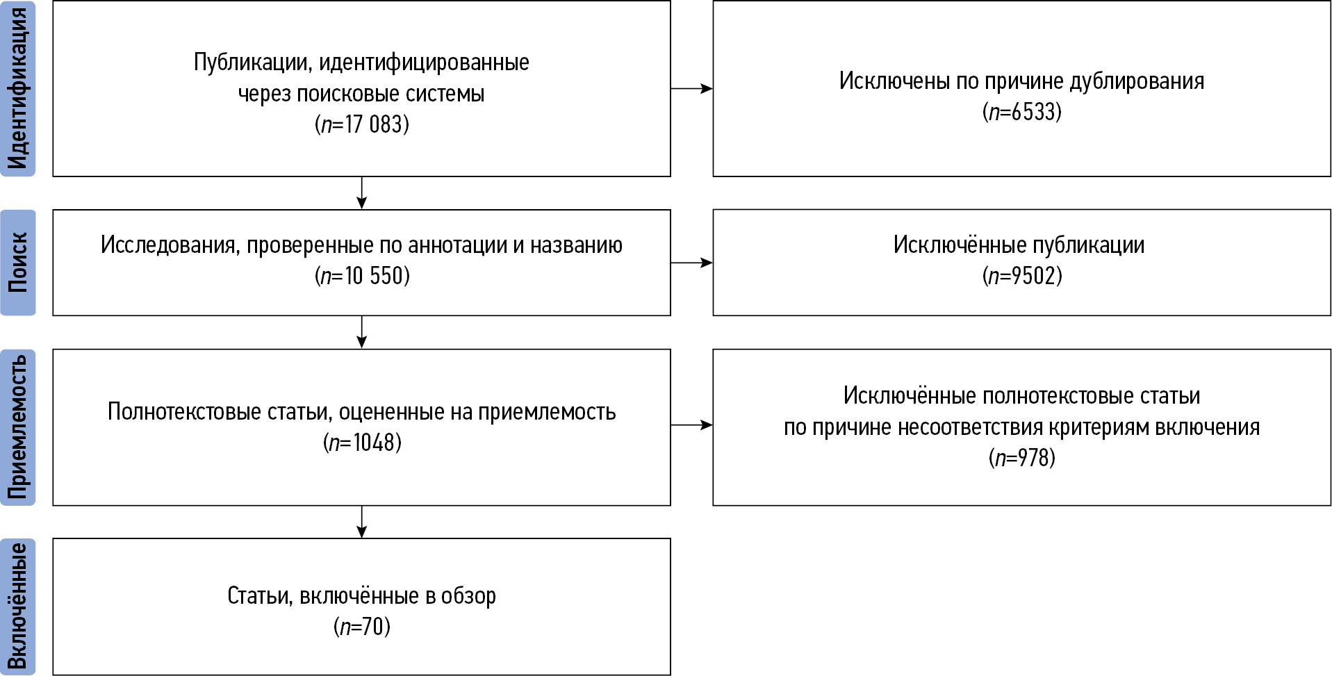
This review summarizes current literature on the application of artificial intelligence technologies in diagnosing cardiovascular diseases and identifies knowledge gaps that require further research. Machine and deep learning methods are widely used and have shown promising results in cardiology. Convolutional neural networks have been used to measure cardiac function parameters from echocardiography results. Deep learning algorithms provide more accurate identification of stenosis and calcification in coronary arteries and characterization of plaques in cardiac CT scans. Convolutional neural networks have been employed for tasks such as automatic segmentation of heart chambers and structures, tissue property determination, and perfusion analysis using magnetic resonance imaging results. As artificial intelligence technologies, particularly machine learning, continue to develop, their integration opens up new possibilities.
Thus, artificial intelligence technologies are of great interest in healthcare, as they enable the rapid analysis of large amounts of data, demonstrating high effectiveness. artificial intelligence can provide additional assistance to specialists, contributing to enhanced workflow efficiency and improved medical care.
Full Text
##article.viewOnOriginalSite##About the authors
Almaz Kh. Islamgulov
Bashkir State Medical University
Author for correspondence.
Email: aslmaz2000@rambler.ru
ORCID iD: 0000-0003-0567-7515
SPIN-code: 8701-3486
Russian Federation, Ufa
Alina S. Bogdanova
Kuban State Medical University
Email: balinochka25@gmail.com
ORCID iD: 0009-0004-9333-5164
Russian Federation, Krasnodar
Damir I. Sufiiarov
Bashkir State Medical University
Email: damur_5@mail.ru
ORCID iD: 0009-0004-3516-6307
SPIN-code: 3311-2947
Russian Federation, Ufa
Alina V. Chernyavskaya
Kuban State Medical University
Email: alinaxxx909@gmail.com
ORCID iD: 0009-0007-8071-1150
Russian Federation, Krasnodar
Elena R. Bairakaeva
Bashkir State Medical University
Email: bairakaeva_0@mail.ru
ORCID iD: 0009-0004-7683-5781
Russian Federation, Ufa
Anastasia A. Maksimova
Bashkir State Medical University
Email: antasiamks@gmail.com
ORCID iD: 0009-0003-4115-2887
Russian Federation, Ufa
Nikita V. Nemychnikov
Bashkir State Medical University
Email: nikita.nemychnikov2001@gmail.com
ORCID iD: 0009-0001-8841-3373
Russian Federation, Ufa
Diana R. Bikieva
Bashkir State Medical University
Email: bikieva.dina@mail.ru
ORCID iD: 0009-0006-5453-5686
SPIN-code: 7078-7424
Russian Federation, Ufa
Alsu I. Shakhmaeva
Bashkir State Medical University
Email: shakhmaeva02@mail.ru
ORCID iD: 0009-0002-8805-9172
Russian Federation, Ufa
Lyubov A. Burdina
Pskov State University
Email: lubovburdina19@gmail.com
ORCID iD: 0009-0004-9199-2515
Russian Federation, Pskov
Aleksandr V. Bolekhan
Pskov State University
Email: sasha-x500@mail.ru
ORCID iD: 0009-0009-3458-2858
Russian Federation, Pskov
Egor I. Akimov
Tula State University
Email: egor.akimov.2001@mail.ru
ORCID iD: 0009-0002-2504-5363
Russian Federation, Tula
Zilya Z. Shurakova
Bashkir State Medical University
Email: divaeva.zilya@mail.ru
ORCID iD: 0009-0007-9625-9787
Russian Federation, Ufa
References
- Kosolapov VP, Yarmonova MV. The analysis of high cardiovascular morbidity and mortality in the adult population as a medical and social problem and the search for ways to solve it. Ural Medical Journal. 2021;20(1):58–64. doi: 10.52420/2071-5943-2021-20-1-58-64 EDN: HCWKUA
- Yeo KK. Artificial intelligence in cardiology: did it take off? Russian Journal for Personalized Medicine. 2023;2(6):16–22. doi: 10.18705/2782-3806-2022-2-6-16-22 EDN: UIENOT
- Xu B, Kocyigit D, Grimm R, et al. Applications of artificial intelligence in multimodality cardiovascular imaging: a state-of-the-art review. Progress in Cardiovascular Diseases. 2020;63(3):367–376. doi: 10.1016/j.pcad.2020.03.003
- Turing AM. I.–Computing machinery and intelligence. Mind. 1950;LIX(236):433–460. doi: 10.1093/mind/LIX.236.433
- McCarthy J, Minsky ML, Rochester N, Shannon CE. A proposal for the dartmouth summer research project on artificial intelligence. AI Mag. 1955;27(4):12. doi: 10.1609/aimag.v27i4.1904
- Komkov AA, Mazaev VP, Ryazanova SV, et al. First study of the RuPatient health information system with optical character recognition of medical records based on machine learning. Cardiovascular Therapy and Prevention. 2022;20(8):91–96. doi: 10.15829/1728-8800-2021-3080 EDN: VOUGRB
- LeCun Y, Bengio Y, Hinton G. Deep learning. Nature. 2015;521:436–444. doi: 10.1038/nature14539
- Gritskov IO, Govorov AV, Vasiliev AO, et al. Data Science – deep learning of neural networks and their application in healthcare. City Healthcare. 2021;2(2):109–115. doi: 10.47619/2713-2617.zm.2021.v2i2;109-115 EDN: SGWBPD
- Vardas PE, Asselbergs FW, van Smeden M, Friedman P. The year in cardiovascular medicine 2021: digital health and innovation. Eur Heart J. 2022;43(4):271–279. doi: 10.1093/eurheartj/ehab874 EDN: CCJAGO
- Maltseva AN, Kop’eva KV, Mochula AV, et al. Association of impaired myocardial flow reserve with risk factors for cardiovascular diseases in patients with nonobstructive coronary artery disease. Russian Journal of Cardiology. 2023;28(2):50–59. (In Russ.) doi: 10.15829/1560-4071-2023-5158 EDN: FNSYNE
- Topol EJ. High-performance medicine: the convergence of human and artificial intelligence. Nat Med. 2019;25(1):44–56. doi: 10.1038/s41591-018-0300-7 EDN: OQSRZW
- Donahue J, Hendricks LA, Rohrbach M, et al. Long-term recurrent convolutional networks for visual recognition and description. IEEE Trans Pattern Anal Mach Intell. 2017;39(4):677–691. doi: 10.1109/TPAMI.2016.2599174
- Sarvamangala DR, Kulkarni RV. Convolutional neural networks in medical image understanding: a survey. Evol Intell. 2022;15(1):1–22. doi: 10.1007/s12065-020-00540-3 EDN: GOCYDD
- Zeleznik R, Foldyna B, Eslami P, et al. Deep convolutional neural networks to predict cardiovascular risk from computed tomography. Nat Commun. 2021;12(1):1–9. doi: 10.1038/s41467-021-20966-2 EDN: KYJQRH
- Amini M, Pursamimi M, Hajianfar G, et al. Machine learning-based diagnosis and risk classification of coronary artery disease using myocardial perfusion imaging SPECT: a radiomics study. Sci Rep. 2023;13(1):14920. doi: 10.1038/s41598-023-42142-w EDN: HGXHIT
- Ambale-Venkatesh B, Yang X, Wu CO, et al. Cardiovascular event prediction by machine learning: the multi-ethnic study of atherosclerosis. Circ Res. 2017;121(9):1092–1101. doi: 10.1161/CIRCRESAHA.117.311312
- Ntalianis E, Cauwenberghs N, Sabovčik F, et al. Feature-based clustering of the left ventricular strain curve for cardiovascular risk stratification in the general population. Front Cardiovasc Med. 2023;10:1263301. doi: 10.3389/fcvm.2023.1263301 EDN: VEPAAS
- Zhao J, Hou X, Pan M, Zhang H. Attention-based generative adversarial network in medical imaging: a narrative review. Comput Biol Med. 2022;149:105948. doi: 10.1016/j.compbiomed.2022.105948 EDN: TBRKVW
- Al'Aref SJ, Maliakal G, Singh G, et al. Machine learning of clinical variables and coronary artery calcium scoring for the prediction of obstructive coronary artery disease on coronary computed tomography angiography: analysis from the CONFIRM registry. Eur Heart J. 2020;41(3):359–367. doi: 10.1093/eurheartj/ehz565 EDN: UYDWAD
- Oikonomou EK, Siddique M, Antoniades C. Artificial intelligence in medical imaging: a radiomic guide to precision phenotyping of cardiovascular disease. Cardiovasc Res. 2020;116(13):2040–2054. doi: 10.1093/cvr/cvaa021 EDN: JJYPCZ
- Fleg JL, Stone GW, Fayad ZA, et al. Detection of high-risk atherosclerotic plaque: report of the NHLBI Working Group on current status and future directions. JACC Cardiovasc Imaging. 2012;5(9):941–955. doi: 10.1016/j.jcmg.2012.07.007
- Chen Q, Zhou F, Xie G, et al. Advances in artificial intelligence-assisted coronary computed tomographic angiography for atherosclerotic plaque characterization. Rev Cardiovasc Med. 2024;25(1):27. doi: 10.31083/j.rcm2501027 EDN: OLVUVT
- Zvartau NE, Solovyova AE, Endubaeva GV, et al. Analysis of the information about the incidence of heart failure, associated mortality and burden on the healthcare system, based on the encoding data in 15 subjects of the Russian Federation. Russian Journal of Cardiology. 2023;28(2S):9–15. doi: 10.15829/1560-4071-2023-5339 EDN: YOUIRD
- Miller PK, Waring L, Bolton GC, Sloane C. Personnel flux and workplace anxiety: personal and interpersonal consequences of understaffing in UK ultrasound departments. Radiography (Lond). 2019;25(1):46–50. doi: 10.1016/j.radi.2018.07.005
- Gao XF, Ge Z, Kong XQ, et al. 3-Year Outcomes of the ULTIMATE Trial Comparing Intravascular Ultrasound Versus Angiography-Guided Drug-Eluting Stent Implantation. JACC Cardiovasc Interv. 2021;14(3):247–257. doi: 10.1016/j.jcin.2020.10.001 EDN: RXYWYL
- Osipova OA, Kontsevaya AV, Demko VV, et al. Elements of artificial intelligence in a predictive personalized model of pharmacotherapy choice in patients with heart failure with mildly reduced ejection fraction of ischemic origin. Cardiovascular Therapy and Prevention. 2023;22(7):16–24. doi: 10.15829/1728-8800-2023-3619 EDN: XLOMXO
- Luong CL, Jafari MH, Behnami D, et al. Validation of machine learning models for estimation of left ventricular ejection fraction on point-of-care ultrasound: insights on features that impact performance. Echo Res Pract. 2024;11(1):9. doi: 10.1186/s44156-024-00043-2
- Olaisen S, Smistad E, Espeland T, et al. Automatic measurements of left ventricular volumes and ejection fraction by artificial intelligence: clinical validation in real time and large databases. Eur Heart J Cardiovasc Imaging. 2024;25(3):383–395. doi: 10.1093/ehjci/jead280 EDN: ALCWDT
- Knackstedt C, Bekkers SC, Schummers G, et al. Fully automated versus standard tracking of left ventricular ejection fraction and longitudinal strain: the FAST-EFs multicenter study. J Am Coll Cardiol. 2015;66(13):1456–1466. doi: 10.1016/j.jacc.2015.07.052
- Narula S, Shameer K, Salem Omar AM, et al. Machine-learning algorithms to automate morphological and functional assessments in 2D echocardiography. J Am Coll Cardiol. 2016;68(21):2287–2295. doi: 10.1016/j.jacc.2016.08.062
- Zhang J, Gajjala S, Agrawal P. Fully automated echocardiogram interpretation in clinical practice. Circulation. 2018;138(16):1623–1635. doi: 10.1161/CIRCULATIONAHA.118.034338
- Sehly A, Jaltotage B, He A. Artificial intelligence in echocardiography: the time is now. Rev Cardiovasc Med. 2022;23(8):256. doi: 10.31083/j.rcm2308256 EDN: LTPRNG
- Playford D, Bordin E, Mohamad R, et al. Enhanced diagnosis of severe aortic stenosis using artificial intelligence: a proof-of-concept study of 530,871 echocardiograms. JACC Cardiovasc Imaging. 2020;13(4):1087–1090. doi: 10.1016/j.jcmg.2019.10.013 EDN: QKVLDF
- Zhang Y, Wang M, Zhang E, Wu Y. Artificial intelligence in the screening, diagnosis, and management of aortic stenosis. Rev Cardiovasc Med. 2024;25(1):31. doi: 10.31083/j.rcm2501031 EDN: MGUQSK
- Ouyang D, He B, Ghorbani A. Video-based AI for beat-to-beat assessment of cardiac function. Nature. 2020;580(7802):252–256. doi: 10.1038/s41586-020-2145-8
- Tereshchenko SN, Zhirov IV, Uskach TM, et al. Eurasian association of cardiology (EAC)/ The National society of heart failure and myocardial disease (NSHFMD) Guidelines for the diagnosis and and treatment of chronic heart failure (2020). Eurasian heart journal. 2020;(3):6–76. doi: 10.38109/2225-1685-2020-3-6-76 EDN: WPQNAB
- Bustin A, Fuin N, Botnar RM, Prieto C. From compressed-sensing to artificial intelligence-based cardiac MRI reconstruction. Front Cardiovasc Med. 2020;7:17. doi: 10.3389/fcvm.2020.00017
- Bai W, Sinclair M, Tarroni G, et al. Automated cardiovascular magnetic resonance image analysis with fully convolutional networks. J Cardiovasc Magn Reson. 2018;20(1):1–12. doi: 10.1186/s12968-018-0471-x EDN: XCDICM
- Bhuva AN, Bai W, Lau C. A multicenter, scan-rescan, human and machine learning CMR study to test generalizability and precision in imaging biomarker analysis. Circ Cardiovasc Imaging. 2019;12(10):e009214. doi: 10.1161/CIRCIMAGING.119.009214
- Celant LR, Wessels JN, Marcus JT. Toward the implementation of optimal cardiac magnetic resonance risk stratification in pulmonary arterial hypertension. Chest. 2024;165(1):181–191. doi: 10.1016/j.chest.2023.07.028 EDN: KJHFYB
- Farrag NA, Lochbihler A, White JA, Ukwatta E. Evaluation of fully automated myocardial segmentation techniques in native and contrast-enhanced T1-mapping cardiovascular magnetic resonance images using fully convolutional neural networks. Med Phys. 2021;48(1):215–226. doi: 10.1002/mp.14574
- Bernard O, Lalande A, Zotti C. Deep learning techniques for automatic MRI cardiac multi-structures segmentation and diagnosis: is the problem solved? IEEE Trans Med Imaging. 2018;37(11):2514–2525. doi: 10.1109/TMI.2018.2837502
- Fahmy AS, Rausch J, Neisius U. Automated cardiac MR scar quantification in hypertrophic cardiomyopathy using deep convolutional neural networks. JACC Cardiovasc Imaging. 2018;11(12):1917–1918. doi: 10.1016/j.jcmg.2018.04.030
- Fahmy AS, Neisius U, Chan RH, et al. Three-dimensional deep convolutional neural networks for automated myocardial scar quantification in hypertrophic cardiomyopathy: a multicenter multivendor study. Radiology. 2020;294(1):52–60. doi: 10.1148/radiol.2019190737
- Chen C, Bai W, Davies RH, et al. Improving the generalizability of convolutional neural network-based segmentation on CMR images. Front Cardiovasc Med. 2020;7:105. doi: 10.3389/fcvm.2020.00105
- Sinitsyn VE, Mershina EA, Larina OM. Cardiac magnetic resonance imaging opportunities in the diagnosis of cardiomyopathy. Clinical and Experimental Surgery. Petrovsky journal. 2014;(1):54–63. EDN: SDUECR
- Khludova LG. Hypersensitivity reactions to contrast media. Astma i allergiya. 2019;(2):8–11. (In Russ.) EDN: GVMUZB
- Zhang Q, Burrage MK, Lukaschuk E, et al. Toward replacing late gadolinium enhancement with artificial intelligence virtual native enhancement for gadolinium-free cardiovascular magnetic resonance tissue characterization in hypertrophic cardiomyopathy. Circulation. 2021;144(8):589–599. doi: 10.1161/CIRCULATIONAHA.121.054432 EDN: BJCVTF
- Leiner T, Rueckert D, Suinesiaputra A, et al. Machine learning in cardiovascular magnetic resonance: basic concepts and applications. J Cardiovasc Magn Reson. 2019;21(1):1–14. doi: 10.1186/s12968-019-0575-y EDN: UTXASC
- Knott KD, Seraphim A, Augusto JB, et al. The prognostic significance of quantitative myocardial perfusion: an artificial intelligence-based approach using perfusion mapping. Circulation. 2020;141(16):1282–1291. doi: 10.1161/CIRCULATIONAHA.119.044666 EDN: HORRTM
- Shesternikova OP, Finn VK, Lesko KA, Vinokurova LV. Intelligent system for predicting the feasibility of using computed tomography. Artificial Intelligence and Decision Making. 2022;(2):3–16. doi: 10.14357/20718594220201 EDN: QSUQRY
- Krittanawong C, Virk HUH, Bangalore S, et al. Machine learning prediction in cardiovascular diseases: a meta-analysis. Sci Rep. 2020;10(1):16057. doi: 10.1038/s41598-020-72685-1 EDN: TUAGSP
- Abdulalimov TP, Obrezan AG. Artificial intelligence capabilities in predicting coronary artery disease. Cardiology: News, Opinions, Training. 2022;10(1):34–39. doi: 10.33029/2309-1908-2022-10-1-34-39 EDN: JRHPMV
- van Rosendael AR, Maliakal G, Kolli KK, et al. Maximization of the usage of coronary CTA derived plaque information using a machine learning based algorithm to improve risk stratification; insights from the CONFIRM registry. J Cardiovasc Comput Tomogr. 2018;12(3):204–209. doi: 10.1016/j.jcct.2018.04.011
- Han D, Kolli KK, Gransar H, et al. Machine learning based risk prediction model for asymptomatic individuals who underwent coronary artery calcium score: comparison with traditional risk prediction approaches. J Cardiovasc Comput Tomogr. 2020;14(2):168–176. doi: 10.1016/j.jcct.2019.09.005
- Motwani M, Dey D, Berman DS, et al. Machine learning for prediction of all-cause mortality in patients with suspected coronary artery disease: a 5-year multicentre prospective registry analysis. Eur Heart J. 2017;38(7):500–507. doi: 10.1093/eurheartj/ehw188
- Klüner LV, Chan K, Antoniades C. Using artificial intelligence to study atherosclerosis from computed tomography imaging: a state-of-the-art review of the current literature. Atherosclerosis. 2024;398:117580. doi: 10.1016/j.atherosclerosis.2024.117580 EDN: BGKJLP
- Oikonomou EK, Marwan M, Desai MY, et al. Non-invasive detection of coronary inflammation using computed tomography and prediction of residual cardiovascular risk (the CRISP CT study): a post-hoc analysis of prospective outcome data. Lancet. 2018;392(10151):929–939. doi: 10.1016/S0140-6736(18)31114-0 EDN: CFUNJT
- Wolterink JM, Leiner T, de Vos BD, et al. Automatic coronary artery calcium scoring in cardiac CT angiography using paired convolutional neural networks. Med Image Anal. 2016;34:123–136. doi: 10.1016/j.media.2016.04.004
- Zreik M, Lessmann N, van Hamersvelt RW, et al. Deep learning analysis of the myocardium in coronary CT angiography for identification of patients with functionally significant coronary artery stenosis. Med Image Anal. 2018;44:72–85. doi: 10.1016/j.media.2017.11.008
- van Hamersvelt RW, Zreik M, Voskuil M, et al. Deep learning analysis of left ventricular myocardium in CT angiographic intermediate-degree coronary stenosis improves the diagnostic accuracy for identification of functionally significant stenosis. Eur Radiol. 2019;29(5):2350–2359. doi: 10.1007/s00330-018-5822-3 EDN: WVYVWW
- Kelm BM, Mittal S, Zheng Y, et al. Detection, grading and classification of coronary stenoses in computed tomography angiography. Med Image Comput Comput Assist Interv. 2011;14(Pt 3):25–32. doi: 10.1007/978-3-642-23626-6_4
- Zreik M, van Hamersvelt RW, Wolterink JM, et al. A recurrent CNN for automatic detection and classification of coronary artery plaque and stenosis in coronary CT angiography. IEEE Trans Med Imaging. 2019;38(7):1588–1598. doi: 10.1109/TMI.2018.2883807
- Bluemke DA, Moy L, Bredella MA, et al. Assessing radiology research on artificial intelligence: a brief guide for authors, reviewers, and readers-from the radiology editorial board. Radiology. 2020;294(3):487–489. doi: 10.1148/radiol.2019192515
- Oakden-Rayner L, Dunnmon J, Carneiro G, Ré C. Hidden stratification causes clinically meaningful failures in machine learning for medical imaging. In: Proc ACM Conf Health Inference Learn (CHIL 2020). Association for Computing Machinery. New York, 2020. P. 151–159. doi: 10.1145/3368555.3384468
- Tjoa E, Guan C. A survey on explainable artificial intelligence (XAI): toward medical XAI. IEEE Trans Neural Netw Learn Syst. 2021;32(11):4793–4813. doi: 10.1109/TNNLS.2020.3027314 EDN: BZXVNY
- DeGrave AJ, Janizek JD, Lee SI. AI for radiographic COVID-19 detection selects shortcuts over signal. Nature Machine Intelligence. 2021;3(7):610–619. doi: 10.1038/s42256-021-00338-7 EDN: MMHUHL
Supplementary files


