

")


腹主动脉分割的计算机断层扫描血管造影数据集
- 作者: Kodenko M.R.1,2, Vasilev Y.A.1, Solovev A.V.1,3, Gatin D.V.1, Yasakova E.P.1, Guseva A.V.1,2, Reshetnikov R.V.1
-
隶属关系:
- Research and Practical Clinical Center for Diagnostics and Telemedicine Technologies
- Bauman Moscow State Technical University
- Morozov Children's City Clinical Hospital
- 期: 卷 6, 编号 1 (2025)
- 页面: 23-32
- 栏目: 数据集
- URL: https://ogarev-online.ru/DD/article/view/310049
- DOI: https://doi.org/10.17816/DD635589
- ID: 310049
如何引用文章

详细
论证。人工智能算法广泛应用于通过不同放射诊断方法获取的图像分析中。这些算法的有效性在很大程度上依赖于相关性和代表性强的训练数据集。当前,迫切需要增加公开数据集的数量,特别是包含计算机断层扫描血管造影腹主动脉图像的数据集,这些数据集不仅包含病变分类信息,还包括血管分割。现有方案的缺点主要包括样本量较小、数据集专一性较强以及准备方法的不一致性。
目的。创建一个开放的数据集,包含计算机断层扫描血管造影图像,并对腹主动脉的正常、扩张、血栓形成及钙化情况进行分割。
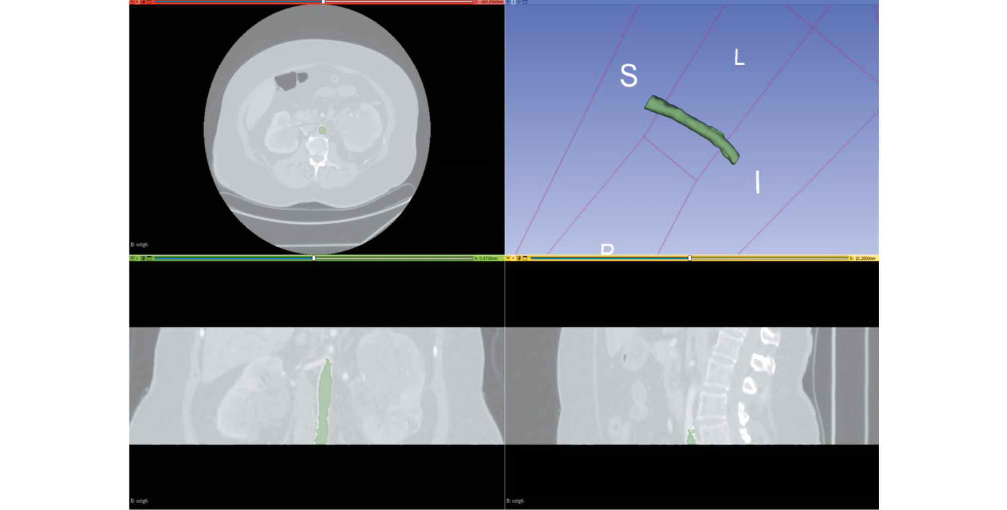
材料与方法。根据人工智能算法测试方法学,制定了数据集准备的技术任务,计算了所需的样本量,并获得了独立伦理委员会的批准。为了创建数据集,采用了之前开发的基于Slicer 3D软件的半自动分割算法。纳入标准:计算机断层扫描血管造影或腹部计算机断层扫描(含造影)结果;具备动脉相扫描;切片厚度 ≤ 3 mm。排除标准:主动脉腔内存在异物;主动脉夹层。该算法在从Unified Radiological Information System中获取的患者研究结果上进行了验证。并对获得的结果与已制定的要求进行了专家评估,同时评估了使用该分割算法所需的时间。
结果。根据要求计算出的样本量为100个包含动脉相扫描且层厚≤1.2 mm的血管造影研究结果。人群数据:独立患者数量为100人,女性患者占比51%;患者年龄中位数为62岁,年龄范围从18岁到84岁不等。61%的病例发现病变(包括联合病变):其中,60个病例表现出钙化迹象,18个病例表现为主动脉扩张,另外18个病例显示有血栓形成迹象。采用分割算法处理一个研究结果(100层扫描)所需的平均时间为0.8小时。
结论。创建了一个包含100个计算机断层扫描血管造影结果的数据集,数据集包含腹主动脉腔的正常、扩张、血栓形成及钙化的分割结果。该数据集已开放,可以用于人工智能算法的开发和测试,以及腹主动脉的类人建模。
作者简介
Maria R. Kodenko
Research and Practical Clinical Center for Diagnostics and Telemedicine Technologies; Bauman Moscow State Technical University
编辑信件的主要联系方式.
Email: m.r.kodenko@yandex.ru
ORCID iD: 0000-0002-0166-3768
SPIN 代码: 5789-0319
Cand. Sci. (Engineering)
俄罗斯联邦, Moscow; MoscowYuriy A. Vasilev
Research and Practical Clinical Center for Diagnostics and Telemedicine Technologies
Email: VasilevYA1@zdrav.mos.ru
ORCID iD: 0000-0002-5283-5961
SPIN 代码: 4458-5608
MD, Cand. Sci. (Medicine)
俄罗斯联邦, MoscowAlexander V. Solovev
Research and Practical Clinical Center for Diagnostics and Telemedicine Technologies; Morozov Children's City Clinical Hospital
Email: SolovevAV10@zdrav.mos.ru
ORCID iD: 0000-0003-4485-2638
SPIN 代码: 9654-4005
俄罗斯联邦, Moscow; Moscow
Denis V. Gatin
Research and Practical Clinical Center for Diagnostics and Telemedicine Technologies
Email: GatinDV@zdrav.mos.ru
ORCID iD: 0000-0002-6218-3012
SPIN 代码: 2256-3564
俄罗斯联邦, Moscow
Elena P. Yasakova
Research and Practical Clinical Center for Diagnostics and Telemedicine Technologies
Email: YasakovaEP@zdrav.mos.ru
ORCID iD: 0000-0003-0315-5502
SPIN 代码: 1047-4692
MD, Cand. Sci. (Medicine)
俄罗斯联邦, MoscowAnastasia V. Guseva
Research and Practical Clinical Center for Diagnostics and Telemedicine Technologies; Bauman Moscow State Technical University
Email: GusevaAV13@zdrav.mos.ru
ORCID iD: 0009-0006-1787-4726
SPIN 代码: 2778-3820
俄罗斯联邦, Moscow; Moscow
Roman V. Reshetnikov
Research and Practical Clinical Center for Diagnostics and Telemedicine Technologies
Email: ReshetnikovRV1@zdrav.mos.ru
ORCID iD: 0000-0002-9661-0254
SPIN 代码: 8592-0558
Cand. Sci. (Physics and Mathematics)
俄罗斯联邦, Moscow参考
- Kumar DS, Bhat V, Gadabanahalli K, Kalyanpur A. Spectrum of abdominal aortic disease in a tertiary health care setup: MDCT based observational study. J Clin Diagn Res. 2016;10(11):TC24–TC29. doi: 10.7860/JCDR/2016/21373.8928
- Russian Society of Angiologists and Vascular Surgeons. Abdominal aortic aneurysm: clinical guidelines [Internet]. Moscow: Russian Society of Angiologists and Vascular Surgeons; 2022 [cited 2022 Apr 7]. (In Russ.) Available from: https://angiolsurgery.org/library/recommendations/2022/aneurysm/recommendation.pdf
- Baliyan V, Shaqdan K, Hedgire S, Ghoshhajra B. Vascular computed tomography angiography technique and indications. Cardiovascular Diagnosis and Therapy. 2019;9(S1):S14S27. doi: 10.21037/CDT.2019.07.04 EDN: IPZHHC
- Alowais ShA, Alghamdi SS, Alsuhebany N, et al. Revolutionizing healthcare: the role of artificial intelligence in clinical practice. BMC Medical Education. 2023;23(1):689. doi: 10.1186/s12909-023-04698-z EDN: AJSDXW
- Ueda D, Kakinuma T, Fujita SH, et al. Fairness of artificial intelligence in healthcare: review and recommendations. Japanese Journal of Radiology. 2023;42(1):3–15. doi: 10.1007/s11604-023-01474-3 EDN: WQQDIA
- Shchupakova AN, Litvyakov AM. Characteristics of atherosclerotic lesion of the abdominal aorta and its unpaired visceral branches in patients with chronic abdominal ischemia. Terapevticheskii arkhiv. 2004;79(6):70–74. EDN: OJZUCJ
- Radl L, Jin YU, Pepe A, et al. AVT: Multicenter aortic vessel tree CTA dataset collection with ground truth segmentation masks. Data in Brief. 2022;40:107801. doi: 10.1016/j.dib.2022.107801 EDN: PEOYKJ
- Imran M, Kreds JR, Gopu VRR, et al. CIS-UNet: Multi-class segmentation of the aorta in computed tomography angiography via context-aware shifted window self-attention. Computerized Medical Imaging and Graphics. 2024;118:102470. doi: 10.1016/j.compmedimag.2024.102470
- Fantazzini A, Esposito M, Finotello A, et al. 3D automatic segmentation of aortic computed tomography angiography combining multi-view 2D convolutional neural networks. Cardiovascular Engineering and Technology. 2020;11(5):576–586. doi: 10.1007/s13239-020-00481-z EDN: FHKUXK
- Jung Y, Kim S, Kim J, et al. Abdominal aortic thrombus segmentation in postoperative computed tomography angiography images using Bi-directional cnvolutional long short-term memory architecture. Sensors. 2022;23(1):175. doi: 10.3390/s23010175 EDN: SGCHXK
- Norgeot B, Quer G, Beaulieu-Jones BK, et al. Minimum information about clinical artificial intelligence modeling: the MI-CLAIM checklist. Nature Medicine. 2020;26(9):1320–1324. doi: 10.1038/s41591-020-1041-y EDN: NRQASJ
- Vasilev YuA, Arzamasov KM, Vladzymyrskyy AV, et al. Preparing a dataset for training and testing software based on artificial intelligence technology: a training manual. Moscow: Moscow Center for Diagnostics and Telemedicine; 2023. (In Russ.) EDN: OGKFGM
- Tymkovich MYu, Avruninn OG, Semenets VV. Using DICOM images in medical systems. Technical Electrodynamics. 2012;(thematic issue):178–183. (In Russ.)
- Li X, Morgan P, Ashburner J, et al. The first step for neuroimaging data analysis: DICOM to NIfTI conversion. Journal of Neuroscience Methods. 2016;264:47–56. doi: 10.1016/j.jneumeth.2016.03.001
- Kodenko MR, Vasilev YuA, Vladzymyrskyy AV. Segmentation of arterial vessels based on CT angiography data using 3D Slicer software: Guidelines. Moscow: Moscow Center for Diagnostics and Telemedicine; 2024. (In Russ.) EDN: CYLZQL
- Kodenko MR, Makarova TA. Preparation of abdominal computed tomography data set for patients with abdominal aortic aneurysm. Digital Diagnostics. 2023;4(1S):90–92. doi: 10.17816/DD430355 EDN: SIUWRL
- Riley RD, Snell KIE, Archer L, et al. Evaluation of clinical prediction models (part 3): calculating the sample size required for an external validation study. BMJ. 2024;384:e074821. doi: 10.1136/bmj-2023-074821
- Hazra A. Using the confidence interval confidently. Journal of Thoracic Disease. 2017;9(10):4124–4129. doi: 10.21037/jtd.2017.09.14
- Vasilev YuA, Vladzymyrskyy AV, Arzamasov KM, et al. Computer vision in radiation diagnostics: the first stage of the Moscow experiment. [Internet]. Moscow: Izdatel'skie resheniya; 2022 [cited 2024 Apr 22]. Available from: https://telemedai.ru/biblioteka-dokumentov/kompyuternoe-zrenie-v-luchevoj-diagnostike-pervyj-etap-moskovskogo-eksperimenta
- Certificate of state registration of the database N 2024621990/ 08.05.2024. Byul. N 5. Vasilev YuА, Kodenko МR, Solovev AV, et al. A computed tomography angiography dataset showing calcification, thrombosis, dilation, and aneurysm, and containing segmentation of the lumen and wall of the abdominal aorta. Available from: https://elibrary.ru/download/elibrary_67262583_69739205.PDF (In Russ.) EDN: FOPTBQ
- Guseva AV, Kodenko MR. Anthropomorphic abdominal aortic phantoms for computed tomography angiography. Digital Diagnostics. 2024;5(1S):27–29. doi: 10.17816/DD626820 EDN: BMDJUN
- Lesage D, Angelini ED, Bloch I, Funka-Lea G. A review of 3D vessel lumen segmentation techniques: models, features and extraction schemes. Medical image analysis. 2009;13(6):819–845. doi: 10.1016/j.media.2009.07.011
补充文件


