

")


Improving aortic aneurysm detection with artificial intelligence based on chest computed tomography data
- Авторлар: Solovev A.V.1,2, Vasilev Y.A.1, Sinitsyn V.E.1,3,4, Petraikin A.V.1, Vladzymyrskyy A.V.1, Shulkin I.M.1, Sharova D.E.1, Semenov D.S.1
-
Мекемелер:
- Research and Practical Clinical Center for Diagnostics and Telemedicine Technologies
- Morozov Children’s Municipal Clinical Hospital
- Clinical City Hospital named after I.V. Davydovsky
- Lomonosov Moscow State University
- Шығарылым: Том 5, № 1 (2024)
- Беттер: 29-40
- Бөлім: Original Study Articles
- URL: https://ogarev-online.ru/DD/article/view/262946
- DOI: https://doi.org/10.17816/DD569388
- ID: 262946
Дәйексөз келтіру

Аннотация
BACKGROUND: Aortic aneurysms are known as “silent killers” because this potentially fatal condition can be asymptomatic. The annual incidence of thoracic aortic aneurysms and ruptures is approximately 10 and 1.6 per 100,000 individuals, respectively. The mortality rate for ruptured aneurysms ranges from 94% to 100%. Early diagnosis and treatment can be life-saving. Artificial intelligence technologies can significantly improve diagnostic accuracy and save the lives of patients with thoracic aortic aneurysms.
AIM: This study aimed to assess the efficacy of artificial intelligence technologies for detecting thoracic aortic aneurysms on chest computed tomography scans, as well as the possibility of using artificial intelligence as a clinical decision support system for radiologists during the primary interpretation of radiological images.
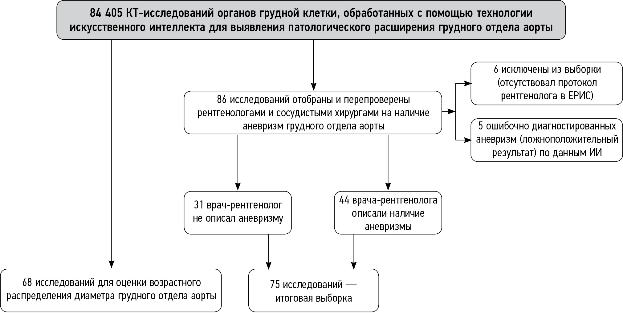
MATERIALS AND METHODS: The results of using artificial intelligence technologies for detecting thoracic aortic aneurysms on non-contrast chest computed tomography scans were evaluated. A sample of 84,405 patients >18 years old was generated, with 86 cases of suspected thoracic aortic aneurysms based on artificial intelligence data selected and retrospectively assessed by radiologists and vascular surgeons. To assess the age distribution of the aortic diameter, an additional sample of 968 cases was randomly selected from the total number.
RESULTS: In 44 cases, aneurysms were initially identified by radiologists, whereas in 31 cases, aneurysms were not detected initially; however, artificial intelligence aided in their detection. Six studies were excluded, and five studies had false-positive results. Artificial intelligence aids in detecting and highlighting aortic pathological changes in medical images, increasing the detection rate of thoracic aortic aneurysms by 41% when interpreting chest computed tomography scans. The use of artificial intelligence technologies for primary interpretations of radiological studies and retrospective assessments is advisable to prevent underdiagnosis of clinically significant pathologies and improve the detection rate of pathological aortic enlargement. In the additional sample, the incidence of thoracic aortic dilation and thoracic aortic aneurysms in adults was 14.5% and 1.2%, respectively. The findings also revealed an age-dependent diameter of the thoracic aorta in both men and women.
CONCLUSION: The use of artificial intelligence technologies in the primary interpretation of chest computed tomography scans can improve the detection rate of clinically significant pathologies such as thoracic aortic aneurysms. Expanding retrospective screening based on chest computed tomography scans using artificial intelligence can improve the diagnosis of concomitant pathologies and prevent negative consequences.
Негізгі сөздер
Толық мәтін
##article.viewOnOriginalSite##Авторлар туралы
Alexander Solovev
Research and Practical Clinical Center for Diagnostics and Telemedicine Technologies; Morozov Children’s Municipal Clinical Hospital
Email: atlantis.92@mail.ru
ORCID iD: 0000-0003-4485-2638
SPIN-код: 9654-4005
Ресей, Moscow; Moscow
Yuriy Vasilev
Research and Practical Clinical Center for Diagnostics and Telemedicine Technologies
Email: VasilevYA1@zdrav.mos.ru
ORCID iD: 0000-0002-0208-5218
SPIN-код: 4458-5608
MD, Cand. Sci. (Medicine)
Ресей, MoscowValentin Sinitsyn
Research and Practical Clinical Center for Diagnostics and Telemedicine Technologies; Clinical City Hospital named after I.V. Davydovsky; Lomonosov Moscow State University
Email: vsini@mail.ru
ORCID iD: 0000-0002-5649-2193
SPIN-код: 8449-6590
MD, Dr. Sci. (Medicine), Professor
Ресей, Moscow; Moscow; MoscowAlexey Petraikin
Research and Practical Clinical Center for Diagnostics and Telemedicine Technologies
Email: atlantis.92@mail.ru
ORCID iD: 0000-0003-1694-4682
SPIN-код: 6193-1656
MD, Dr. Sci. (Medicine)
Ресей, MoscowAnton Vladzymyrskyy
Research and Practical Clinical Center for Diagnostics and Telemedicine Technologies
Email: VladzimirskijAV@zdrav.mos.ru
ORCID iD: 0000-0002-2990-7736
SPIN-код: 3602-7120
MD, Dr. Sci. (Medicine)
Ресей, MoscowIgor Shulkin
Research and Practical Clinical Center for Diagnostics and Telemedicine Technologies
Email: ShulkinIM@zdrav.mos.ru
ORCID iD: 0000-0002-7613-5273
SPIN-код: 5266-0618
Ресей, Moscow
Daria Sharova
Research and Practical Clinical Center for Diagnostics and Telemedicine Technologies
Email: SharovaDE@zdrav.mos.ru
ORCID iD: 0000-0001-5792-3912
SPIN-код: 1811-7595
Ресей, Moscow
Dmitry Semenov
Research and Practical Clinical Center for Diagnostics and Telemedicine Technologies
Хат алмасуға жауапты Автор.
Email: SemenovDS4@zdrav.mos.ru
ORCID iD: 0000-0002-4293-2514
SPIN-код: 2278-7290
Cand. Sci. (Engineering)
Ресей, MoscowӘдебиет тізімі
- The top 10 causes of death [Internet]. World Health Organization. [cited 12 May 2023]. Available from: https://www.who.int/ru/news-room/fact-sheets/detail/the-top-10-causes-of-death
- Gouveia e Melo R, Silva Duarte G, Lopes A, et al. Incidence and Prevalence of Thoracic Aortic Aneurysms: A Systematic Review and Meta-analysis of Population-Based Studies. Seminars in thoracic and cardiovascular surgery. 2022;34(1):1–16. doi: 10.1053/j.semtcvs.2021.02.029
- Clinical guidelines. Guidelines for the diagnosis and treatment of aortic diseases (2017). Russian Journal of Cardiology and Cardiovascular Surgery. 2018;11(1):7–67. EDN: YPAKRP
- Kuznechevsky FV, Osipov AKh, Evsikov EM, Abramov IS, Otarova SM. Prevalence and clinical features of aorta aneurysm; and dissections: 10-year results of consequent autopsies made at O.M. Filatov city clinical hospital №15. Russian Journal of Cardiology. 2004;9(6):5–13. EDN: ISVRYL
- Irtyuga OB, Voronkina IV, Smagina LV, et al. The frequency to detect of ascending aorta aneurysms and the mechanism of its development according register of the Almazov Federal Heart, Blood and Endocrinology Centre. Bulletin of Almazov Federal Heart, Blood and Endocrinology Centre. 2011;(5):73–78. EDN: OWGHOB
- Lavall D, Schäfers HJ, Böhm M, Laufs U. Aneurysms of the ascending aorta. Deutsches Arzteblatt international. 2012;109(13):227–233. doi: 10.3238/arztebl.2012.0227
- Olsson C, Thelin S, Ståhle E, Ekbom A, Granath F. Thoracic Aortic Aneurysm and Dissection. Circulation. 2006;114(24):2611–2618. doi: 10.1161/CIRCULATIONAHA.106.630400
- Elefteriades JA. Natural history of thoracic aortic aneurysms: indications for surgery, and surgical versus nonsurgical risks. The Annals of thoracic surgery. 2002;74(5):1877–1880. doi: 10.1016/s0003-4975(02)04147-4
- Tsai EB, Chiles C, Carter BW, et al. Incidental Findings on Lung Cancer Screening: Significance and Management. Seminars in ultrasound, CT, and MR. 2018;39(3):273–281. doi: 10.1053/j.sult.2018.02.005
- Chernina VYu, Blokhin IA, Nikolaev AE, et al. Tactics for the management of incidentalomas. Section 3. Thyroid, pituitary, vasculature and mediastinum. Moscow: Research and Practical Clinical Center for Diagnostics and Telemedicine; 2019. (In Russ). EDN: WSYSYP
- Law M. “Opportunistic” Screening. J Med Screen. 1994;1(4):208. doi: 10.1177/096914139400100403
- Kumar Y, Hooda K, Li S, et al. Abdominal aortic aneurysm: pictorial review of common appearances and complications. Annals of translational medicine. 2017;5(12):256. doi: 10.21037/atm.2017.04.32
- Vasilev YuA, Vladzymyrskyy AV, editors. Computer Vision in Radiologic Diagnostics: the First Stage of the Moscow Experiment. Moscow: Limited Liability Company Izdatelskie reshenia; 2022. (In Russ). EDN: FOYLXK
- Erbel R, Aboyans V, Boileau C, Vlachopoulos C. 2014 ESC Guidelines on the diagnosis and treatment of aortic diseases: Document covering acute and chronic aortic diseases of the thoracic and abdominal aorta of the adult. The Task Force for the Diagnosis and Treatment of Aortic Diseases of the European. European heart journal. 2014;35(41):2873–2926. doi: 10.1093/eurheartj/ehu281
- Documents on the Experiment [Internet]. Center for Diagnostics and Telemedicine. [cited 16 June 2023]. Available from: https://mosmed.ai/ai/docs/
- Chest-IRA [Internet]. Center for Diagnostics and Telemedicine. [cited 16 June 2023]. Available from: https://mosmed.ai/service_catalog/chestira/
- Evangelista A, Sitges M, Jondeau G, et al. Multimodality imaging in thoracic aortic diseases: a clinical consensus statement from the European Association of Cardiovascular Imaging and the European Society of Cardiology working group on aorta and peripheral vascular diseases. European Heart Journal Cardiovascular Imaging. 2023;24(5):e65–e85. doi: 10.1093/ehjci/jead024
- Etli M, Avnioglu S, Yilmaz H, Karahan O. Investigation of the correlation between cardiac parameters and aortic diameter in patients with ascending aortic aneurysm. Egyptian Heart Journal. 2022;74(1):1–7. doi: 10.1186/s43044-022-00238-0
- Pearce W, Slaughter M, Lemaire S, et al. Aortic diameter as a function of age, gender, and body surface area. Surgery. 1993;114(4):691–697.
- Vasilev YA, Bobrovskaya TM, Arzamasov KM, et al. Medical datasets for machine learning: fundamental principles of standartization and systematization. Manager Zdravoohranenia. 2023;4:28–41. EDN: EPGAMD doi: 10.21045/1811-0185-2023-4-28-41
- Chetverikov SF, Arzamasov KM, Andreichenko AE, et al. Approaches to Sampling for Quality Control of Artificial Intelligence in Biomedical Research. Modern Technologies in Medicine. 2023;15(2):19–25. EDN: FUKXYC doi: 10.17691/stm2023.15.2.02
- Zinchenko VV, Arzamasov KM, Chetverikov SF, et al. Methodology for Conducting Post-Marketing Surveillance of Software as a Medical Device Based on Artificial Intelligence Technologies. Modern Technologies in Medicine. 2022;14(5):15–25. doi: 10.17691/stm2022.14.5.02
- Chernina VY, Belyaev MG, Silin AY, et al. A diagnostic and economic evaluation of the complex artificial intelligence algorithm aimed to detect 10 pathologies on the chest CT images. medRxiv. 2023;4. doi: 10.1101/2023.04.19.23288584
- Macruz FBC, Lu C, Strout J, et al. Quantification of the Thoracic Aorta and Detection of Aneurysm at CT: Development and Validation of a Fully Automatic Methodology. Radiology: Artificial Intelligence. 2022;4(2):e210076. doi: 10.1148/ryai.210076
- Adam C, Fabre D, Mougin J, et al. Pre-surgical and Post-surgical Aortic Aneurysm Maximum Diameter Measurement: Full Automation by Artificial Intelligence. European Journal of Vascular and Endovascular Surgery. 2021;62(6):869–877. doi: 10.1016/j.ejvs.2021.07.013
- Vladzymyrskyy AV, Kudryavtsev ND, Kozhikhina DD, et al. Effectiveness of using artificial intelligence technologies for dual descriptions of the results of preventive lung examinations. Profilakticheskaya Meditsina. 2022;25(7):7–15. doi: 10.17116/profmed2022250717
- Rodriguez-Ruiz A, Lång K, Gubern-Merida A, et al. Stand-Alone Artificial Intelligence for Breast Cancer Detection in Mammography: Comparison With 101 Radiologists. Journal of the National Cancer Institute. 2019;111(9):916–922. doi: 10.1093/jnci/djy222
- Rueckel J, Reidler P, Fink N, et al. Artificial intelligence assistance improves reporting efficiency of thoracic aortic aneurysm CT follow-up. European journal of radiology. 2021;134(134):109424. doi: 10.1016/j.ejrad.2020.109424
- Tang A, Tam R, Cadrin-Chênevert A, et al. Canadian Association of Radiologists White Paper on Artificial Intelligence in Radiology. Canadian Association of Radiologists journal. 2018;69(2):120–135. doi: 10.1016/j.carj.2018.02.002
- Certificate of state registration of the database № 2023621046/ 30.03.2023. Vasilev YuA, Turavilova EV, Shul’kin IM, et al. MosMedData: CT scan with signs of abdominal aortic aneurysm. (In Russ). EDN: LXROHZ
- Aliev AF, Kudryavtsev ND, Petraikin AV, et al. Changing of pulmonary artery diameter in accordance with severity of COVID-19 (assessment based on non-contrast computer tomography). Digital Diagnostics. 2021;2(3):249–260. EDN: VTMKCJ doi: 10.17816/DD76726
- Morozov SP, Shapieva AN, Narkevich BYa, et al. Informativity of radial diagnostics methods in various pathological conditions of the organism. Moscow: Research and Practical Clinical Center for Diagnostics and Telemedicine; 2020. (In Russ). EDN: DYEYBT
- Vasilev YuA, Vladzymyrskyy AV, Bondarchuk DV, et al. Importance of artificial intelligence technologies to prevent defects in radiologist’s practice. Medical doctor and IT. 2023;(2):16–27. EDN: SYZAOQ doi: 10.25881/18110193_2023_2_16
Қосымша файлдар


